


Structure and function studies on Helicobacter pylori arginase
Zhang, J., Zhang, X., Li, D., Hu, Y., Zou, Q., Wang, D.To be published.
Experimental Data Snapshot
wwPDB Validation 3D Report Full Report

Entity ID: 1 | |||||
|---|---|---|---|---|---|
| Molecule | Chains | Sequence Length | Organism | Details | Image |
| Arginase (RocF) | 330 | Helicobacter pylori 26695 | Mutation(s): 0 Gene Names: HP_1399 | 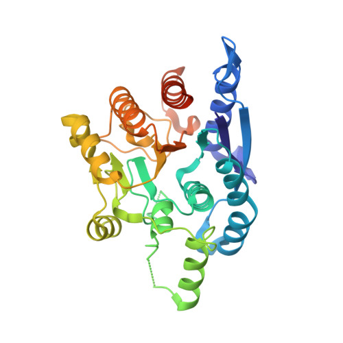 | |
UniProt | |||||
Find proteins for O25949 (Helicobacter pylori (strain ATCC 700392 / 26695)) Explore O25949 Go to UniProtKB: O25949 | |||||
Entity Groups | |||||
| Sequence Clusters | 30% Identity50% Identity70% Identity90% Identity95% Identity100% Identity | ||||
| UniProt Group | O25949 | ||||
Sequence AnnotationsExpand | |||||
| |||||
| Ligands 1 Unique | |||||
|---|---|---|---|---|---|
| ID | Chains | Name / Formula / InChI Key | 2D Diagram | 3D Interactions | |
| MN Query on MN | E [auth A] F [auth A] G [auth B] H [auth B] I [auth C] | MANGANESE (II) ION Mn WAEMQWOKJMHJLA-UHFFFAOYSA-N |  | ||
| Length ( Å ) | Angle ( ˚ ) |
|---|---|
| a = 94.69 | α = 90 |
| b = 102.24 | β = 90 |
| c = 148.61 | γ = 90 |
| Software Name | Purpose |
|---|---|
| HKL-2000 | data collection |
| PHASES | phasing |
| CNS | refinement |
| DENZO | data reduction |
| SCALA | data scaling |
RCSB PDB (citation) is hosted by
RCSB PDB is a member of the

