4G11

X-ray structure of PI3K-gamma bound to a 4-(morpholin-4-yl)- (6-oxo-1,6-dihydropyrimidin-2-yl)amide inhibitor


Experimental Data Snapshot

  • Method: X-RAY DIFFRACTION
  • Resolution: 3.40 Å
  • R-Value Free: 0.349 
  • R-Value Work: 0.282 
  • R-Value Observed: 0.285 

wwPDB Validation   3D Report Full Report


Ligand Structure Quality Assessment 


This is version 1.1 of the entry. See complete history


Literature

Preparation and optimization of new 4-(morpholin-4-yl)-(6-oxo-1,6-dihydropyrimidin-2-yl)amide derivatives as PI3K beta inhibitors

Certal, V.Halley, F.Virone-Oddos, A.Thompson, F.Filoche-Romme, B.El-Ahmad, Y.Carry, J.C.Delorme, C.Karlsson, A.Abecassis, P.Y.Vincent, L.Bonnevaux, H.Nicolas, J.P.Morales, R.Michot, N.Vade, I.Louboutin, A.Perron, S.Doerflinger, G.Tric, B.Monget, S.Lengauer, C.Schio, L.

(2012) Bioorg Med Chem Lett 22: 6381-6384

  • DOI: https://doi.org/10.1016/j.bmcl.2012.08.072
  • Primary Citation of Related Structures:  
    4G11

  • PubMed Abstract: 

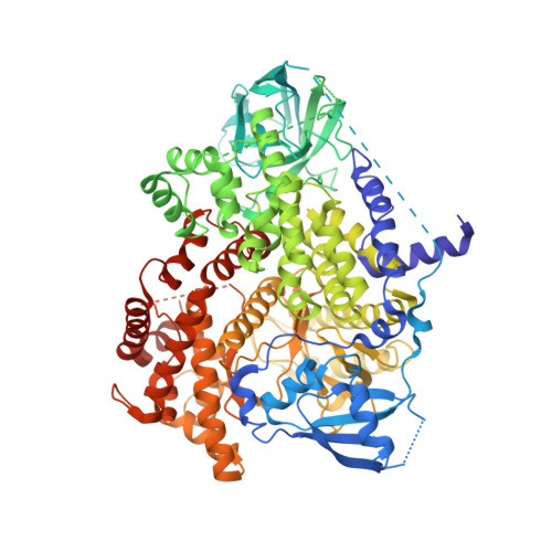
    From a HTS campaign, a new series of pyrimidone anilides exemplified by compound 1 has been identified with good inhibitory activity for the PI3Kβ isoform. The structure of compound 1 in PI3Kγ was solved revealing a binding mode in agreement with the SAR observed on PI3Kβ. These compounds displayed inhibition in the nanomolar range in the biochemical assay and were also potent p-Akt inhibitors in a PTEN-deficient PC3 prostate cancer cell line. Optimization of in vitro pharmocokinetic properties led to compound 25 exhibiting 52% bioavailability in mice and target engagement in an acute PK/PD study.


  • Organizational Affiliation

    Medicinal Chemistry Oncology Drug Discovery, Sanofi, Vitry-sur-Seine, France.


Macromolecules
Find similar proteins by:  (by identity cutoff)  |  3D Structure
Entity ID: 1
MoleculeChains Sequence LengthOrganismDetailsImage
Phosphatidylinositol 4,5-bisphosphate 3-kinase catalytic subunit gamma isoform966Homo sapiensMutation(s): 0 
Gene Names: PIK3CG
EC: 2.7.1.153 (PDB Primary Data), 2.7.11.1 (PDB Primary Data)
UniProt & NIH Common Fund Data Resources
Find proteins for P48736 (Homo sapiens)
Explore P48736 
Go to UniProtKB:  P48736
PHAROS:  P48736
GTEx:  ENSG00000105851 
Entity Groups  
Sequence Clusters30% Identity50% Identity70% Identity90% Identity95% Identity100% Identity
UniProt GroupP48736
Sequence Annotations
Expand
  • Reference Sequence
Small Molecules
Ligands 1 Unique
IDChains Name / Formula / InChI Key2D Diagram3D Interactions
0W7
Query on 0W7

Download Ideal Coordinates CCD File 
B [auth A]2-[4-(morpholin-4-yl)-6-oxo-1,6-dihydropyrimidin-2-yl]-N-phenylacetamide
C16 H18 N4 O3
FPFCFOIMTFHSNY-UHFFFAOYSA-N
Binding Affinity Annotations 
IDSourceBinding Affinity
0W7 BindingDB:  4G11 IC50: 1.00e+4 (nM) from 1 assay(s)
PDBBind:  4G11 IC50: 1.00e+4 (nM) from 1 assay(s)
Experimental Data & Validation

Experimental Data

  • Method: X-RAY DIFFRACTION
  • Resolution: 3.40 Å
  • R-Value Free: 0.349 
  • R-Value Work: 0.282 
  • R-Value Observed: 0.285 
  • Space Group: C 1 2 1
Unit Cell:
Length ( Å )Angle ( ˚ )
a = 140.63α = 90
b = 67.38β = 96.14
c = 105.89γ = 90
Software Package:
Software NamePurpose
DNAdata collection
MOLREPphasing
REFMACrefinement
PROCESSdata reduction
SCALAdata scaling

Structure Validation

View Full Validation Report


Ligand Structure Quality Assessment 


Entry History 

Deposition Data

  • Released Date: 2012-11-14 
  • Deposition Author(s): Morales, R.

Revision History  (Full details and data files)

  • Version 1.0: 2012-11-14
    Type: Initial release
  • Version 1.1: 2023-09-13
    Changes: Data collection, Database references, Derived calculations, Refinement description