4FXQ

Full-length Certhrax toxin from Bacillus cereus in complex with Inhibitor P6


Experimental Data Snapshot

  • Method: X-RAY DIFFRACTION
  • Resolution: 1.96 Å
  • R-Value Free: 0.227 
  • R-Value Work: 0.187 
  • R-Value Observed: 0.189 

wwPDB Validation   3D Report Full Report


Ligand Structure Quality Assessment 


This is version 1.4 of the entry. See complete history


Literature

Certhrax Toxin, an Anthrax-related ADP-ribosyltransferase from Bacillus cereus.

Visschedyk, D.Rochon, A.Tempel, W.Dimov, S.Park, H.W.Merrill, A.R.

(2012) J Biol Chem 287: 41089-41102

  • DOI: https://doi.org/10.1074/jbc.M112.412809
  • Primary Citation of Related Structures:  
    4FK7, 4FXQ, 4GF1

  • PubMed Abstract: 

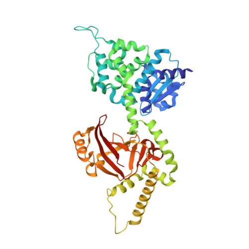
    We identified Certhrax, the first anthrax-like mART toxin from the pathogenic G9241 strain of Bacillus cereus. Certhrax shares 31% sequence identity with anthrax lethal factor from Bacillus anthracis; however, we have shown that the toxicity of Certhrax resides in the mART domain, whereas anthrax uses a metalloprotease mechanism. Like anthrax lethal factor, Certhrax was found to require protective antigen for host cell entry. This two-domain enzyme was shown to be 60-fold more toxic to mammalian cells than anthrax lethal factor. Certhrax localizes to distinct regions within mouse RAW264.7 cells by 10 min postinfection and is extranuclear in its cellular location. Substitution of catalytic residues shows that the mART function is responsible for the toxicity, and it binds NAD(+) with high affinity (K(D) = 52.3 ± 12.2 μM). We report the 2.2 Å Certhrax structure, highlighting its structural similarities and differences with anthrax lethal factor. We also determined the crystal structures of two good inhibitors (P6 (K(D) = 1.7 ± 0.2 μM, K(i) = 1.8 ± 0.4 μM) and PJ34 (K(D) = 5.8 ± 2.6 μM, K(i) = 9.6 ± 0.3 μM)) in complex with Certhrax. As with other toxins in this family, the phosphate-nicotinamide loop moves toward the NAD(+) binding site with bound inhibitor. These results indicate that Certhrax may be important in the pathogenesis of B. cereus.


  • Organizational Affiliation

    Department of Molecular and Cellular Biology, University of Guelph, Guelph, Ontario N1G 2W1, Canada.


Macromolecules
Find similar proteins by:  (by identity cutoff)  |  3D Structure
Entity ID: 1
MoleculeChains Sequence LengthOrganismDetailsImage
Putative ADP-ribosyltransferase Certhrax
A, B
460Bacillus cereusMutation(s): 0 
Gene Names: Amino acids 18-471BCE_G9241_pBC218_0027pBC218_0027
EC: 2.4.2
UniProt
Find proteins for Q4MV79 (Bacillus cereus)
Explore Q4MV79 
Go to UniProtKB:  Q4MV79
Entity Groups  
Sequence Clusters30% Identity50% Identity70% Identity90% Identity95% Identity100% Identity
UniProt GroupQ4MV79
Sequence Annotations
Expand
  • Reference Sequence
Small Molecules
Binding Affinity Annotations 
IDSourceBinding Affinity
G9L PDBBind:  4FXQ Kd: 1700 (nM) from 1 assay(s)
Binding MOAD:  4FXQ Kd: 1700 (nM) from 1 assay(s)
Experimental Data & Validation

Experimental Data

  • Method: X-RAY DIFFRACTION
  • Resolution: 1.96 Å
  • R-Value Free: 0.227 
  • R-Value Work: 0.187 
  • R-Value Observed: 0.189 
  • Space Group: P 21 21 21
Unit Cell:
Length ( Å )Angle ( ˚ )
a = 61.31α = 90
b = 100.29β = 90
c = 191.09γ = 90
Software Package:
Software NamePurpose
MxDCdata collection
PHASERphasing
PHENIXmodel building
PHENIXrefinement
AutoProcessdata reduction
BESTdata reduction
pointlessdata scaling
XDSdata reduction
HKL-2000data reduction
iMOSFLMdata reduction
SCALAdata scaling
PHENIXphasing

Structure Validation

View Full Validation Report


Ligand Structure Quality Assessment 


Entry History 

Deposition Data

Revision History  (Full details and data files)

  • Version 1.0: 2012-09-26
    Type: Initial release
  • Version 1.1: 2012-10-03
    Changes: Database references
  • Version 1.2: 2012-12-19
    Changes: Database references
  • Version 1.3: 2024-02-28
    Changes: Data collection, Database references, Derived calculations
  • Version 1.4: 2024-04-03
    Changes: Refinement description