4FRZ

Arabidopsis KCBP motor domain dimerized via regulatory domain


Experimental Data Snapshot

  • Method: X-RAY DIFFRACTION
  • Resolution: 2.40 Å
  • R-Value Work: 0.225 
  • R-Value Observed: 0.263 

wwPDB Validation   3D Report Full Report


Ligand Structure Quality Assessment 


This is version 1.1 of the entry. See complete history


Literature

Plant Kinesin-Like Calmodulin Binding Protein Employs Its Regulatory Domain for Dimerization.

Vinogradova, M.V.Malanina, G.G.Waitzman, J.S.Rice, S.E.Fletterick, R.J.

(2013) PLoS One 8: e66669-e66669

  • DOI: https://doi.org/10.1371/journal.pone.0066669
  • Primary Citation of Related Structures:  
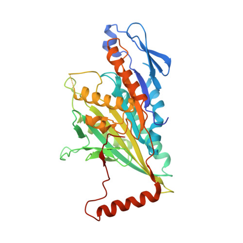
    4FRZ

  • PubMed Abstract: 

    Kinesin-like calmodulin binding protein (KCBP), a Kinesin-14 family motor protein, is involved in the structural organization of microtubules during mitosis and trichome morphogenesis in plants. The molecular mechanism of microtubule bundling by KCBP remains unknown. KCBP binding to microtubules is regulated by Ca(2+)-binding proteins that recognize its C-terminal regulatory domain. In this work, we have discovered a new function of the regulatory domain. We present a crystal structure of an Arabidopsis KCBP fragment showing that the C-terminal regulatory domain forms a dimerization interface for KCBP. This dimerization site is distinct from the dimerization interface within the N-terminal domain. Side chains of hydrophobic residues of the calmodulin binding helix of the regulatory domain form the C-terminal dimerization interface. Biochemical experiments show that another segment of the regulatory domain located beyond the dimerization interface, its negatively charged coil, is unexpectedly and absolutely required to stabilize the dimers. The strong microtubule bundling properties of KCBP are unaffected by deletion of the C-terminal regulatory domain. The slow minus-end directed motility of KCBP is also unchanged in vitro. Although the C-terminal domain is not essential for microtubule bundling, we suggest that KCBP may use its two independent dimerization interfaces to support different types of bundled microtubule structures in cells. Two distinct dimerization sites may provide a mechanism for microtubule rearrangement in response to Ca(2+) signaling since Ca(2+)- binding proteins can disengage KCBP dimers dependent on its C-terminal dimerization interface.


  • Organizational Affiliation

    Department of Biochemistry and Biophysics, University of California San Francisco, San Francisco, California, United States of America.


Macromolecules
Find similar proteins by:  (by identity cutoff)  |  3D Structure
Entity ID: 1
MoleculeChains Sequence LengthOrganismDetailsImage
Kinesin-like calmodulin-binding protein
A, B
386Arabidopsis thalianaMutation(s): 1 
Gene Names: KCBPZWIAt5g65930K14B20.10
UniProt
Find proteins for Q9FHN8 (Arabidopsis thaliana)
Explore Q9FHN8 
Go to UniProtKB:  Q9FHN8
Entity Groups  
Sequence Clusters30% Identity50% Identity70% Identity90% Identity95% Identity100% Identity
UniProt GroupQ9FHN8
Sequence Annotations
Expand
  • Reference Sequence
Small Molecules
Ligands 4 Unique
IDChains Name / Formula / InChI Key2D Diagram3D Interactions
ADP
Query on ADP

Download Ideal Coordinates CCD File 
D [auth A],
J [auth B]
ADENOSINE-5'-DIPHOSPHATE
C10 H15 N5 O10 P2
XTWYTFMLZFPYCI-KQYNXXCUSA-N
IMD
Query on IMD

Download Ideal Coordinates CCD File 
E [auth A],
F [auth A],
G [auth A],
K [auth B]
IMIDAZOLE
C3 H5 N2
RAXXELZNTBOGNW-UHFFFAOYSA-O
EDO
Query on EDO

Download Ideal Coordinates CCD File 
H [auth A],
L [auth B],
M [auth B]
1,2-ETHANEDIOL
C2 H6 O2
LYCAIKOWRPUZTN-UHFFFAOYSA-N
MG
Query on MG

Download Ideal Coordinates CCD File 
C [auth A],
I [auth B]
MAGNESIUM ION
Mg
JLVVSXFLKOJNIY-UHFFFAOYSA-N
Experimental Data & Validation

Experimental Data

  • Method: X-RAY DIFFRACTION
  • Resolution: 2.40 Å
  • R-Value Work: 0.225 
  • R-Value Observed: 0.263 
  • Space Group: P 1 21 1
Unit Cell:
Length ( Å )Angle ( ˚ )
a = 45.713α = 90
b = 75.068β = 91.45
c = 120.598γ = 90
Software Package:
Software NamePurpose
ADSCdata collection
CNSrefinement
HKL-2000data reduction
HKL-2000data scaling
CNSphasing

Structure Validation

View Full Validation Report


Ligand Structure Quality Assessment 


Entry History 

Deposition Data

Revision History  (Full details and data files)

  • Version 1.0: 2013-07-10
    Type: Initial release
  • Version 1.1: 2023-09-13
    Changes: Data collection, Database references, Derived calculations, Refinement description