


4FR5
Crystal Structure of Shikimate Dehydrogenase (aroE) Y210S Mutant from Helicobacter pylori in Complex with Shikimate
- PDB DOI: https://doi.org/10.2210/pdb4FR5/pdb
- Classification: OXIDOREDUCTASE
- Organism(s): Helicobacter pylori 26695
- Expression System: Escherichia coli BL21(DE3)
- Mutation(s): Yes
- Deposited: 2012-06-26 Released: 2012-12-26
Experimental Data Snapshot
- Method: X-RAY DIFFRACTION
- Resolution: 2.20 Å
- R-Value Free: 0.231
- R-Value Work: 0.195
- R-Value Observed: 0.197

This is version 1.1 of the entry. See complete history.

Macromolecules
Find similar proteins by:
(by identity cutoff) | 3D Structure
Entity ID: 1 | |||||
|---|---|---|---|---|---|
| Molecule | Chains | Sequence Length | Organism | Details | Image |
| Shikimate dehydrogenase | 271 | Helicobacter pylori 26695 | Mutation(s): 1 Gene Names: aroE, HP_1249 EC: 1.1.1.25 | 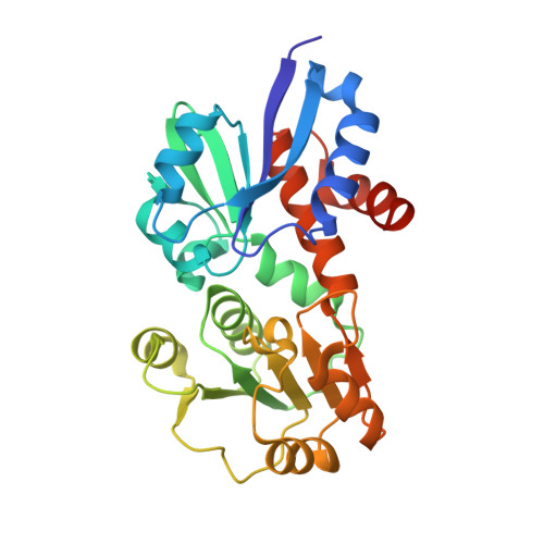 | |
UniProt | |||||
Find proteins for P56119 (Helicobacter pylori (strain ATCC 700392 / 26695)) Explore P56119 Go to UniProtKB: P56119 | |||||
Entity Groups | |||||
| Sequence Clusters | 30% Identity50% Identity70% Identity90% Identity95% Identity100% Identity | ||||
| UniProt Group | P56119 | ||||
Sequence AnnotationsExpand | |||||
| |||||
Small Molecules
| Ligands 1 Unique | |||||
|---|---|---|---|---|---|
| ID | Chains | Name / Formula / InChI Key | 2D Diagram | 3D Interactions | |
| SKM Query on SKM | C [auth A], D [auth B] | (3R,4S,5R)-3,4,5-TRIHYDROXYCYCLOHEX-1-ENE-1-CARBOXYLIC ACID C7 H10 O5 JXOHGGNKMLTUBP-HSUXUTPPSA-N |  | ||
Experimental Data & Validation
Experimental Data
- Method: X-RAY DIFFRACTION
- Resolution: 2.20 Å
- R-Value Free: 0.231
- R-Value Work: 0.195
- R-Value Observed: 0.197
- Space Group: P 21 21 21
Unit Cell:
| Length ( Å ) | Angle ( ˚ ) |
|---|---|
| a = 46.507 | α = 90 |
| b = 88.523 | β = 90 |
| c = 118.185 | γ = 90 |
| Software Name | Purpose |
|---|---|
| REFMAC | refinement |
| PDB_EXTRACT | data extraction |
Entry History
Deposition Data
- Released Date: 2012-12-26 Deposition Author(s): Cheng, W.C., Chen, T.J., Wang, W.C.
Revision History (Full details and data files)
- Version 1.0: 2012-12-26
Type: Initial release - Version 1.1: 2023-09-13
Changes: Data collection, Database references, Derived calculations, Refinement description
