4FB2

Crystal Structure of Substrate-Free P450cin


Experimental Data Snapshot

  • Method: X-RAY DIFFRACTION
  • Resolution: 1.37 Å
  • R-Value Free: 0.167 
  • R-Value Work: 0.136 
  • R-Value Observed: 0.138 

wwPDB Validation   3D Report Full Report


Ligand Structure Quality Assessment 


This is version 1.4 of the entry. See complete history


Literature

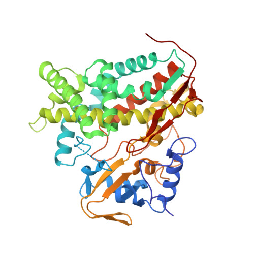
Crystal structures of substrate-free and nitrosyl cytochrome p450cin: implications for o(2) activation.

Madrona, Y.Tripathi, S.Li, H.Poulos, T.L.

(2012) Biochemistry 51: 6623-6631

  • DOI: https://doi.org/10.1021/bi300666u
  • Primary Citation of Related Structures:  
    4FB2, 4FMX, 4FYZ, 4G3R

  • PubMed Abstract: 

    The crystal structure of the P450cin substrate-bound nitric oxide complex and the substrate-free form have been determined revealing a substrate-free structure that adopts an open conformation relative to the substrate-bound structure. The region of the I helix that forms part of the O(2) binding pocket shifts from an α helix in the substrate-free form to a π helix in the substrate-bound form. Unique to P450cin is an active site residue, Asn242, in the I helix that H-bonds with the substrate. In most other P450s this residue is a Thr and plays an important role in O(2) activation by participating in an H-bonding network required for O(2) activation. The π/α I helix transition results in the carbonyl O atom of Gly238 moving in to form an H-bond with the water/hydroxide ligand in the substrate-free form. The corresponding residue, Gly248, in the substrate-free P450cam structure experiences a similar motion. Most significantly, in the oxy-P450cam complex Gly248 adopts a position midway between the substrate-free and -bound states. A comparison between these P450cam and the new P450cin structures provides insights into differences in how the two P450s activate O(2). The structure of P450cin complexed with nitric oxide, a close mimic of the O(2) complex, shows that Gly238 is likely to form tighter interactions with ligands than the corresponding Gly248 in P450cam. Having a close interaction between an H-bond acceptor, the Gly238 carbonyl O atom, and the distal oxygen atom of O(2) will promote protonation and hence further reduction of the oxy complex to the hydroperoxy intermediate resulting in heterolytic cleavage of the peroxide O-O bond and formation of the active ferryl intermediate required for substrate hydroxylation.


  • Organizational Affiliation

    Department of Molecular Biology and Biochemistry, University of California, Irvine, CA 92697-3900, USA.


Macromolecules
Find similar proteins by:  (by identity cutoff)  |  3D Structure
Entity ID: 1
MoleculeChains Sequence LengthOrganismDetailsImage
P450cin
A, B, C, D
398Citrobacter braakiiMutation(s): 0 
Gene Names: cinA
UniProt
Find proteins for Q8VQF6 (Citrobacter braakii)
Explore Q8VQF6 
Go to UniProtKB:  Q8VQF6
Entity Groups  
Sequence Clusters30% Identity50% Identity70% Identity90% Identity95% Identity100% Identity
UniProt GroupQ8VQF6
Sequence Annotations
Expand
  • Reference Sequence
Small Molecules
Ligands 3 Unique
IDChains Name / Formula / InChI Key2D Diagram3D Interactions
HEM
Query on HEM

Download Ideal Coordinates CCD File 
E [auth A],
H [auth B],
K [auth C],
P [auth D]
PROTOPORPHYRIN IX CONTAINING FE
C34 H32 Fe N4 O4
KABFMIBPWCXCRK-RGGAHWMASA-L
EDO
Query on EDO

Download Ideal Coordinates CCD File 
F [auth A],
I [auth B],
L [auth C],
M [auth C],
O [auth C]
1,2-ETHANEDIOL
C2 H6 O2
LYCAIKOWRPUZTN-UHFFFAOYSA-N
CL
Query on CL

Download Ideal Coordinates CCD File 
G [auth A],
J [auth B],
N [auth C],
Q [auth D]
CHLORIDE ION
Cl
VEXZGXHMUGYJMC-UHFFFAOYSA-M
Experimental Data & Validation

Experimental Data

  • Method: X-RAY DIFFRACTION
  • Resolution: 1.37 Å
  • R-Value Free: 0.167 
  • R-Value Work: 0.136 
  • R-Value Observed: 0.138 
  • Space Group: P 1
Unit Cell:
Length ( Å )Angle ( ˚ )
a = 60.383α = 96.81
b = 83.912β = 96.39
c = 88.192γ = 89.94
Software Package:
Software NamePurpose
HKL-2000data collection
PHASERphasing
PHENIXrefinement
HKL-2000data reduction
SCALEPACKdata scaling

Structure Validation

View Full Validation Report


Ligand Structure Quality Assessment 


Entry History 

Deposition Data

Revision History  (Full details and data files)

  • Version 1.0: 2012-07-18
    Type: Initial release
  • Version 1.1: 2012-08-22
    Changes: Database references
  • Version 1.2: 2012-09-05
    Changes: Database references
  • Version 1.3: 2014-05-28
    Changes: Data collection
  • Version 1.4: 2023-09-13
    Changes: Data collection, Database references, Derived calculations, Refinement description