4FAY

Crystal structure of a trimeric bacterial microcompartment shell protein PduB with glycerol metabolites


Experimental Data Snapshot

  • Method: X-RAY DIFFRACTION
  • Resolution: 1.56 Å
  • R-Value Free: 0.202 
  • R-Value Work: 0.173 
  • R-Value Observed: 0.174 

wwPDB Validation   3D Report Full Report


This is version 1.2 of the entry. See complete history


Literature

Substrate channels revealed in the trimeric Lactobacillus reuteri bacterial microcompartment shell protein PduB.

Pang, A.Liang, M.Prentice, M.B.Pickersgill, R.W.

(2012) Acta Crystallogr D Biol Crystallogr 68: 1642-1652

  • DOI: https://doi.org/10.1107/S0907444912039315
  • Primary Citation of Related Structures:  
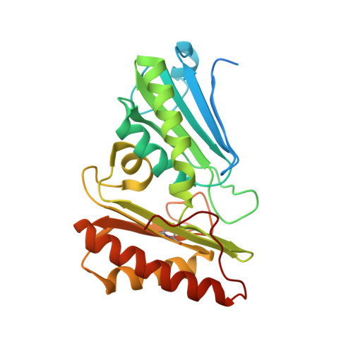
    4FAY

  • PubMed Abstract: 

    Lactobacillus reuteri metabolizes two similar three-carbon molecules, 1,2-propanediol and glycerol, within closed polyhedral subcellular bacterial organelles called bacterial microcompartments (metabolosomes). The outer shell of the propanediol-utilization (Pdu) metabolosome is composed of hundreds of mainly hexagonal protein complexes made from six types of protein subunits that share similar domain structures. The structure of the bacterial microcompartment protein PduB has a tandem structural repeat within the subunit and assembles into a trimer with pseudo-hexagonal symmetry. This trimeric structure forms sheets in the crystal lattice and is able to fit within a polymeric sheet of the major shell component PduA to assemble a facet of the polyhedron. There are three pores within the trimer and these are formed between the tandem repeats within the subunits. The structure shows that each of these pores contains three glycerol molecules that interact with conserved residues, strongly suggesting that these subunit pores channel glycerol substrate into the metabolosome. In addition to the observation of glycerol occupying the subunit channels, the presence of glycerol on the molecular threefold symmetry axis suggests a role in locking closed the central region.


  • Organizational Affiliation

    School of Biological and Chemical Sciences, Queen Mary University of London, 327 Mile End Road, London, England.


Macromolecules
Find similar proteins by:  (by identity cutoff)  |  3D Structure
Entity ID: 1
MoleculeChains Sequence LengthOrganismDetailsImage
Microcompartments protein
A, B, C
258Limosilactobacillus reuteri subsp. reuteriMutation(s): 0 
Gene Names: Lreu_1748PduB
UniProt
Find proteins for A5VMB3 (Limosilactobacillus reuteri (strain DSM 20016))
Explore A5VMB3 
Go to UniProtKB:  A5VMB3
Entity Groups  
Sequence Clusters30% Identity50% Identity70% Identity90% Identity95% Identity100% Identity
UniProt GroupA5VMB3
Sequence Annotations
Expand
  • Reference Sequence
Small Molecules
Ligands 2 Unique
IDChains Name / Formula / InChI Key2D Diagram3D Interactions
GOL
Query on GOL

Download Ideal Coordinates CCD File 
D [auth A]
E [auth A]
F [auth A]
G [auth A]
H [auth A]
D [auth A],
E [auth A],
F [auth A],
G [auth A],
H [auth A],
I [auth A],
J [auth A],
K [auth B],
L [auth B],
M [auth B],
N [auth B],
O [auth B],
P [auth B],
R [auth C],
S [auth C],
T [auth C],
U [auth C]
GLYCEROL
C3 H8 O3
PEDCQBHIVMGVHV-UHFFFAOYSA-N
ACT
Query on ACT

Download Ideal Coordinates CCD File 
Q [auth B]ACETATE ION
C2 H3 O2
QTBSBXVTEAMEQO-UHFFFAOYSA-M
Experimental Data & Validation

Experimental Data

  • Method: X-RAY DIFFRACTION
  • Resolution: 1.56 Å
  • R-Value Free: 0.202 
  • R-Value Work: 0.173 
  • R-Value Observed: 0.174 
  • Space Group: C 2 2 21
Unit Cell:
Length ( Å )Angle ( ˚ )
a = 69.865α = 90
b = 120.864β = 90
c = 145.531γ = 90
Software Package:
Software NamePurpose
ADSCdata collection
PHENIXmodel building
REFMACrefinement
XDSdata reduction
SCALAdata scaling
PHENIXphasing

Structure Validation

View Full Validation Report


Entry History 

Deposition Data

Revision History  (Full details and data files)

  • Version 1.0: 2012-11-21
    Type: Initial release
  • Version 1.1: 2012-12-12
    Changes: Database references
  • Version 1.2: 2024-04-03
    Changes: Data collection, Database references, Derived calculations, Refinement description