4FAS

Complex crystal structure of hydroxylamine oxidoreductase and NE1300 from Nitrosomonas europaea


Experimental Data Snapshot

  • Method: X-RAY DIFFRACTION
  • Resolution: 2.10 Å
  • R-Value Free: 0.206 
  • R-Value Work: 0.165 
  • R-Value Observed: 0.167 

wwPDB Validation   3D Report Full Report


Ligand Structure Quality Assessment 


This is version 1.3 of the entry. See complete history


Literature

Structural studies of hydroxylamine oxidoreductase reveal a unique heme cofactor and a previously unidentified interaction partner.

Cedervall, P.E.Hooper, A.B.Wilmot, C.M.

(2013) Biochemistry 52: 6211-6218

  • DOI: https://doi.org/10.1021/bi400960w
  • Primary Citation of Related Structures:  
    4FAS

  • PubMed Abstract: 

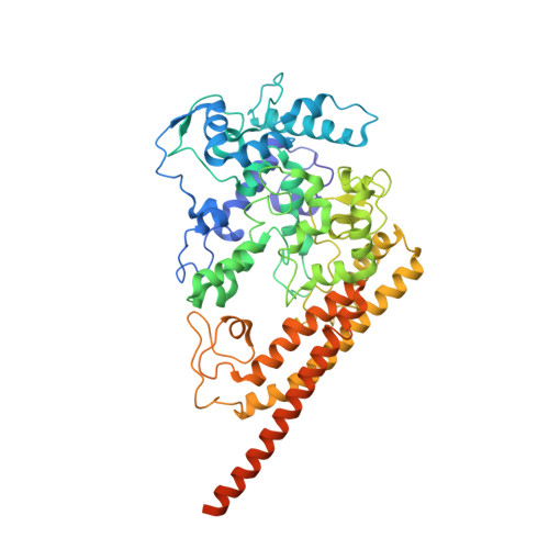
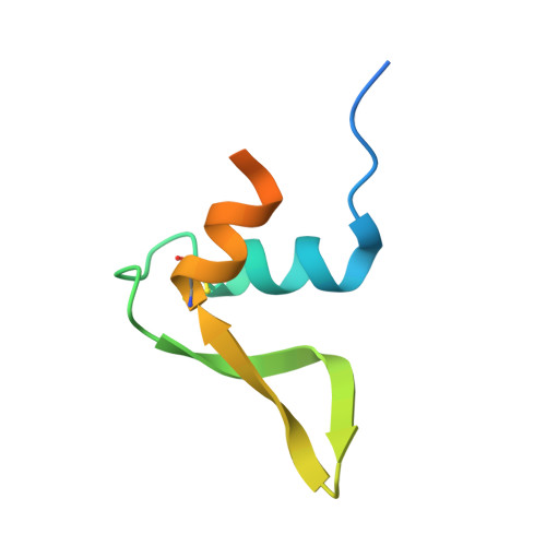
    Hydroxylamine oxidoreductase (HAO) is a 24-heme homotrimeric enzyme that catalyzes the conversion of hydroxylamine to nitrite in nitrifying bacteria: a key reaction in the nitrogen cycle. One heme in each HAO monomer is a highly unusual heme P460 that is the site of catalysis. This was proposed to be a c-type heme that contained an additional porphyrin-tyrosine cross-link. Here, we report the crystal structure of HAO from Nitrosomonas europaea to 2.1 Å resolution that defines a different model compatible with the crystallographic and biochemical data. The structure reveals that heme P460 contains two covalent cross-links between the porphyrin and a Tyr residue. In addition, the enzyme was purified from source, and an unknown physiological HAO binding partner was present within the crystal (annotated in the genome as hypothetical protein NE1300). NE1300 may play a structural role in the ternary complex with cytochrome c554, the physiological electron acceptor of HAO.


  • Organizational Affiliation

    Department of Biochemistry, Molecular Biology, and Biophysics, University of Minnesota , Minneapolis, Minnesota 55455, United States.


Macromolecules
Find similar proteins by:  (by identity cutoff)  |  3D Structure
Entity ID: 1
MoleculeChains Sequence LengthOrganismDetailsImage
Hydroxylamine oxidoreductase
A, B, C
546Nitrosomonas europaea ATCC 19718Mutation(s): 0 
EC: 1.7.3.4
UniProt
Find proteins for Q50925 (Nitrosomonas europaea (strain ATCC 19718 / CIP 103999 / KCTC 2705 / NBRC 14298))
Explore Q50925 
Go to UniProtKB:  Q50925
Entity Groups  
Sequence Clusters30% Identity50% Identity70% Identity90% Identity95% Identity100% Identity
UniProt GroupQ50925
Sequence Annotations
Expand
  • Reference Sequence
Find similar proteins by:  (by identity cutoff)  |  3D Structure
Entity ID: 2
MoleculeChains Sequence LengthOrganismDetailsImage
NE1300
D, E, F
69Nitrosomonas europaea ATCC 19718Mutation(s): 0 
UniProt
Find proteins for Q82V11 (Nitrosomonas europaea (strain ATCC 19718 / CIP 103999 / KCTC 2705 / NBRC 14298))
Explore Q82V11 
Go to UniProtKB:  Q82V11
Entity Groups  
Sequence Clusters30% Identity50% Identity70% Identity90% Identity95% Identity100% Identity
UniProt GroupQ82V11
Sequence Annotations
Expand
  • Reference Sequence
Small Molecules
Ligands 8 Unique
IDChains Name / Formula / InChI Key2D Diagram3D Interactions
ISW
Query on ISW

Download Ideal Coordinates CCD File 
GA [auth B],
N [auth A],
UA [auth C]
{3,3'-[(9S)-8,13-diethenyl-3,7,12,17-tetramethyl-9,10-dihydroporphyrin-2,18-diyl-kappa~4~N~21~,N~22~,N~23~,N~24~]dipropanoato(2-)}iron
C34 H34 Fe N4 O4
QSWMAEGFLATCMU-JMAPEOGHSA-L
HEC
Query on HEC

Download Ideal Coordinates CCD File 
AA [auth B]
AB [auth C]
BA [auth B]
BB [auth C]
CA [auth B]
AA [auth B],
AB [auth C],
BA [auth B],
BB [auth C],
CA [auth B],
DA [auth B],
EA [auth B],
FA [auth B],
G [auth A],
H [auth A],
I [auth A],
J [auth A],
K [auth A],
L [auth A],
M [auth A],
VA [auth C],
WA [auth C],
XA [auth C],
YA [auth C],
Z [auth B],
ZA [auth C]
HEME C
C34 H34 Fe N4 O4
HXQIYSLZKNYNMH-LJNAALQVSA-N
P6G
Query on P6G

Download Ideal Coordinates CCD File 
LA [auth B]HEXAETHYLENE GLYCOL
C12 H26 O7
IIRDTKBZINWQAW-UHFFFAOYSA-N
PG4
Query on PG4

Download Ideal Coordinates CCD File 
MA [auth B]TETRAETHYLENE GLYCOL
C8 H18 O5
UWHCKJMYHZGTIT-UHFFFAOYSA-N
PGE
Query on PGE

Download Ideal Coordinates CCD File 
HB [auth C],
U [auth A]
TRIETHYLENE GLYCOL
C6 H14 O4
ZIBGPFATKBEMQZ-UHFFFAOYSA-N
PEG
Query on PEG

Download Ideal Coordinates CCD File 
CB [auth C]
DB [auth C]
EB [auth C]
FB [auth C]
GB [auth C]
CB [auth C],
DB [auth C],
EB [auth C],
FB [auth C],
GB [auth C],
HA [auth B],
IA [auth B],
JA [auth B],
KA [auth B],
O [auth A],
P [auth A],
PB [auth C],
Q [auth A],
R [auth A],
RB [auth F],
S [auth A],
T [auth A]
DI(HYDROXYETHYL)ETHER
C4 H10 O3
MTHSVFCYNBDYFN-UHFFFAOYSA-N
EDO
Query on EDO

Download Ideal Coordinates CCD File 
IB [auth C]
JB [auth C]
KB [auth C]
LB [auth C]
NA [auth B]
IB [auth C],
JB [auth C],
KB [auth C],
LB [auth C],
NA [auth B],
NB [auth C],
OA [auth B],
OB [auth C],
RA [auth B],
SA [auth B],
TA [auth B],
V [auth A],
W [auth A],
X [auth A],
Y [auth A]
1,2-ETHANEDIOL
C2 H6 O2
LYCAIKOWRPUZTN-UHFFFAOYSA-N
NO3
Query on NO3

Download Ideal Coordinates CCD File 
MB [auth C],
PA [auth B],
QA [auth B],
QB [auth F]
NITRATE ION
N O3
NHNBFGGVMKEFGY-UHFFFAOYSA-N
Experimental Data & Validation

Experimental Data

  • Method: X-RAY DIFFRACTION
  • Resolution: 2.10 Å
  • R-Value Free: 0.206 
  • R-Value Work: 0.165 
  • R-Value Observed: 0.167 
  • Space Group: P 21 21 2
Unit Cell:
Length ( Å )Angle ( ˚ )
a = 140.731α = 90
b = 142.618β = 90
c = 107.376γ = 90
Software Package:
Software NamePurpose
SBC-Collectdata collection
PHASERphasing
REFMACrefinement
HKL-2000data reduction
HKL-2000data scaling

Structure Validation

View Full Validation Report


Ligand Structure Quality Assessment 


Entry History 

Deposition Data

Revision History  (Full details and data files)

  • Version 1.0: 2013-09-04
    Type: Initial release
  • Version 1.1: 2013-11-20
    Changes: Database references
  • Version 1.2: 2017-11-15
    Changes: Refinement description
  • Version 1.3: 2023-09-13
    Changes: Data collection, Database references, Derived calculations, Refinement description