4F3J

Crystal Structure of Trimeric gC1q Domain of Human C1QTNF5 associated with Late-onset Retinal Macular Degeneration


Experimental Data Snapshot

  • Method: X-RAY DIFFRACTION
  • Resolution: 1.34 Å
  • R-Value Free: 0.165 
  • R-Value Work: 0.136 
  • R-Value Observed: 0.138 

wwPDB Validation   3D Report Full Report


This is version 1.2 of the entry. See complete history


Literature

Crystal structure of the globular domain of C1QTNF5: Implications for late-onset retinal macular degeneration.

Tu, X.Palczewski, K.

(2012) J Struct Biol 180: 439-446

  • DOI: https://doi.org/10.1016/j.jsb.2012.07.011
  • Primary Citation of Related Structures:  
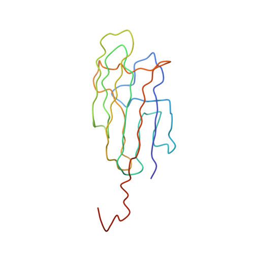
    4F3J

  • PubMed Abstract: 

    Autosomal dominant late-onset retinal macular degeneration (L-ORMD) is caused by a single S163R mutation in the C1q and tumor necrosis factor-related protein 5 (C1QTNF5) gene. The C1QTNF5 gene encodes a secreted and membrane-associated protein involved in adhesion of retinal pigmented epithelial cells (RPE) to Bruch's membrane. The crystal structure of the trimeric globular domain of human C1QTNF5 at 1.34Å resolution reveals unique features of this novel C1q family member. It lacks a Ca²⁺-binding site, displays a remarkable non-uniform distribution of surface electrostatic potentials and possesses a unique sequence (F₁₈₁F₁₈₂G₁₈₃G₁₈₄W₁₈₅P₁₈₆) that forms a hydrophobic plateau surrounded by Lys and Arg residues with a solvent cavity underneath. S₁₆₃ forms a hydrogen bond with F₁₈₂ in a hydrophobic area extending to the hydrophobic plateau. The pathogenic mutation S163R disrupts this hydrogen bonding and positively charges these hydrophobic areas. Thus, our analysis provides insights into the structural basis of the L-ORMD disease mechanism.


  • Organizational Affiliation

    Department of Pharmacology, School of Medicine, Case Western Reserve University, OH, USA.


Macromolecules
Find similar proteins by:  (by identity cutoff)  |  3D Structure
Entity ID: 1
MoleculeChains Sequence LengthOrganismDetailsImage
Complement C1q tumor necrosis factor-related protein 5148Homo sapiensMutation(s): 0 
Gene Names: C1QTNF5CTRP5UNQ303/PRO344
UniProt & NIH Common Fund Data Resources
Find proteins for Q9BXJ0 (Homo sapiens)
Explore Q9BXJ0 
Go to UniProtKB:  Q9BXJ0
PHAROS:  Q9BXJ0
GTEx:  ENSG00000223953 
Entity Groups  
Sequence Clusters30% Identity50% Identity70% Identity90% Identity95% Identity100% Identity
UniProt GroupQ9BXJ0
Sequence Annotations
Expand
  • Reference Sequence
Experimental Data & Validation

Experimental Data

  • Method: X-RAY DIFFRACTION
  • Resolution: 1.34 Å
  • R-Value Free: 0.165 
  • R-Value Work: 0.136 
  • R-Value Observed: 0.138 
  • Space Group: H 3
Unit Cell:
Length ( Å )Angle ( ˚ )
a = 46.621α = 90
b = 46.621β = 90
c = 138.83γ = 120
Software Package:
Software NamePurpose
SCALAdata scaling
PHASERphasing
PHENIXrefinement
PDB_EXTRACTdata extraction
ADSCdata collection
XSCALEdata scaling

Structure Validation

View Full Validation Report


Entry History 

Deposition Data

Revision History  (Full details and data files)

  • Version 1.0: 2012-11-07
    Type: Initial release
  • Version 1.1: 2012-12-12
    Changes: Database references
  • Version 1.2: 2024-02-28
    Changes: Data collection, Database references