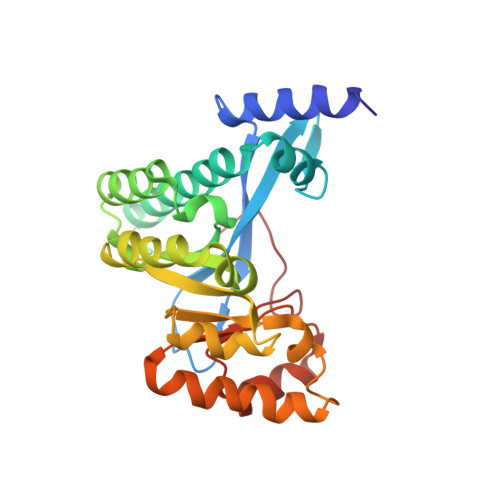
4F3H

The structural of FimXEAL-c-di-GMP from Xanthomonas campestris


Experimental Data Snapshot

  • Method: X-RAY DIFFRACTION
  • Resolution: 2.50 Å
  • R-Value Free: 0.259 
  • R-Value Work: 0.234 
  • R-Value Observed: 0.234 

wwPDB Validation   3D Report Full Report


Ligand Structure Quality Assessment 


This is version 1.2 of the entry. See complete history


Literature

Structural polymorphism of c-di-GMP bound to an EAL domain and in complex with a type II PilZ-domain protein.

Chin, K.-H.Kuo, W.-T.Yu, Y.-J.Liao, Y.-T.Yang, M.T.Chou, S.-H.

(2012) Acta Crystallogr D Biol Crystallogr 68: 1380-1392

  • DOI: https://doi.org/10.1107/S0907444912030594
  • Primary Citation of Related Structures:  
    4F3H, 4F48

  • PubMed Abstract: 

    Cyclic di-GMP (c-di-GMP) is a novel secondary-messenger molecule that is involved in regulating a plethora of important bacterial activities through binding to an unprecedented array of effectors. Proteins with a canonical PilZ domain that bind c-di-GMP play crucial roles in regulating flagellum-based motility. In contrast, noncanonical type II PilZ domains that do not effectively bind c-di-GMP regulate twitching motility, which is dependent on type IV pili (T4P). Recent data indicate that T4P biogenesis is initiated via the interaction of a noncanonical type II PilZ protein with the GGDEF/EAL-domain protein FimX and the pilus motor protein PilB at high c-di-GMP concentrations. However, the molecular details of such interactions remain to be elucidated. In this manuscript, the first hetero-complex crystal structure between a type II PilZ protein and the EAL domain of the FimX protein (FimX(EAL)) from Xanthomonas campestris pv. campestris (Xcc) in the presence of c-di-GMP is reported. This work reveals two novel conformations of monomeric c-di-GMP in the XccFimX(EAL)-c-di-GMP and XccFimX(EAL)-c-di-GMP-XccPilZ complexes, as well as a unique interaction mode of a type II PilZ domain with FimX(EAL). These findings indicate that c-di-GMP is sufficiently flexible to adjust its conformation to match the corresponding recognition motifs of different cognate effectors. Together, these results represent a first step towards an understanding of how T4P biogenesis is controlled by c-di-GMP at the molecular level and also of the ability of c-di-GMP to bind to a wide variety of effectors.


  • Organizational Affiliation

    Agricultural Biotechnology Center, National Chung Hsing University, Taichung 40227, Taiwan.


Macromolecules
Find similar proteins by:  (by identity cutoff)  |  3D Structure
Entity ID: 1
MoleculeChains Sequence LengthOrganismDetailsImage
Putative uncharacterized protein250Xanthomonas campestris pv. campestrisMutation(s): 0 
Gene Names: XCC2291
UniProt
Find proteins for Q8P8F1 (Xanthomonas campestris pv. campestris (strain ATCC 33913 / DSM 3586 / NCPPB 528 / LMG 568 / P 25))
Explore Q8P8F1 
Go to UniProtKB:  Q8P8F1
Entity Groups  
Sequence Clusters30% Identity50% Identity70% Identity90% Identity95% Identity100% Identity
UniProt GroupQ8P8F1
Sequence Annotations
Expand
  • Reference Sequence
Small Molecules
Ligands 1 Unique
IDChains Name / Formula / InChI Key2D Diagram3D Interactions
C2E
Query on C2E

Download Ideal Coordinates CCD File 
B [auth A]9,9'-[(2R,3R,3aS,5S,7aR,9R,10R,10aS,12S,14aR)-3,5,10,12-tetrahydroxy-5,12-dioxidooctahydro-2H,7H-difuro[3,2-d:3',2'-j][1,3,7,9,2,8]tetraoxadiphosphacyclododecine-2,9-diyl]bis(2-amino-1,9-dihydro-6H-purin-6-one)
C20 H24 N10 O14 P2
PKFDLKSEZWEFGL-MHARETSRSA-N
Binding Affinity Annotations 
IDSourceBinding Affinity
C2E PDBBind:  4F3H Kd: 420 (nM) from 1 assay(s)
Experimental Data & Validation

Experimental Data

  • Method: X-RAY DIFFRACTION
  • Resolution: 2.50 Å
  • R-Value Free: 0.259 
  • R-Value Work: 0.234 
  • R-Value Observed: 0.234 
  • Space Group: P 32 2 1
Unit Cell:
Length ( Å )Angle ( ˚ )
a = 65.665α = 90
b = 65.665β = 90
c = 121.29γ = 120
Software Package:
Software NamePurpose
HKL-2000data collection
CNSrefinement
HKL-2000data reduction
HKL-2000data scaling
CNSphasing

Structure Validation

View Full Validation Report


Ligand Structure Quality Assessment 


Entry History 

Deposition Data

Revision History  (Full details and data files)

  • Version 1.0: 2012-11-14
    Type: Initial release
  • Version 1.1: 2019-12-25
    Changes: Database references
  • Version 1.2: 2023-11-08
    Changes: Data collection, Database references, Derived calculations, Refinement description, Structure summary