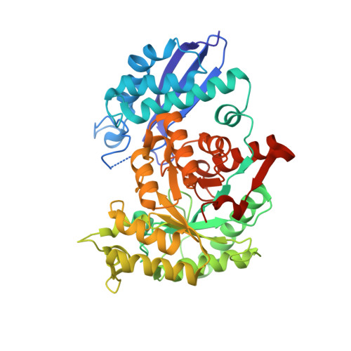
An octamer of enolase from Streptococcus suis.
Lu, Q., Lu, H., Qi, J., Lu, G., Gao, G.F.(2012) Protein Cell 3: 769-780
- PubMed: 23055041
- DOI: https://doi.org/10.1007/s13238-012-2040-7
- Primary Citation of Related Structures:
4EWJ - PubMed Abstract:
Enolase is a conserved cytoplasmic metalloenzyme existing universally in both eukaryotic and prokaryotic cells. The enzyme can also locate on the cell surface and bind to plasminogen, via which contributing to the mucosal surface localization of the bacterial pathogens and assisting the invasion into the host cells. The functions of the eukaryotic enzymes on the cell surface expression (including T cells, B cells, neutrophils, monocytoes, neuronal cells and epithelial cells) are not known. Streptococcus suis serotype 2 (S. suis 2, SS2) is an important zoonotic pathogen which has recently caused two large-scale outbreaks in southern China with severe streptococcal toxic shock syndrome (STSS) never seen before in human sufferers. We recently identified the SS2 enolase as an important protective antigen which could protect mice from fatal S.suis 2 infection. In this study, a 2.4-angstrom structure of the SS2 enolase is solved, revealing an octameric arrangement in the crystal. We further demonstrated that the enzyme exists exclusively as an octamer in solution via a sedimentation assay. These results indicate that the octamer is the biological unit of SS2 enolase at least in vitro and most likely in vivo as well. This is, to our knowledge, the first comprehensive characterization of the SS2 enolase octamer both structurally and biophysically, and the second octamer enolase structure in addition to that of Streptococcus pneumoniae. We also investigated the plasminogen binding property of the SS2 enzyme.
Organizational Affiliation:
CAS Key Laboratory of Pathogenic Microbiology and Immunology, Institute of Microbiology, Chinese Academy of Sciences, Beijing, China.