4ELT

Snapshot of the large fragment of DNA polymerase I from Thermus Aquaticus processing modified pyrimidines


Experimental Data Snapshot

  • Method: X-RAY DIFFRACTION
  • Resolution: 2.20 Å
  • R-Value Free: 0.209 
  • R-Value Work: 0.160 
  • R-Value Observed: 0.163 

wwPDB Validation   3D Report Full Report


Ligand Structure Quality Assessment 


This is version 1.1 of the entry. See complete history


Literature

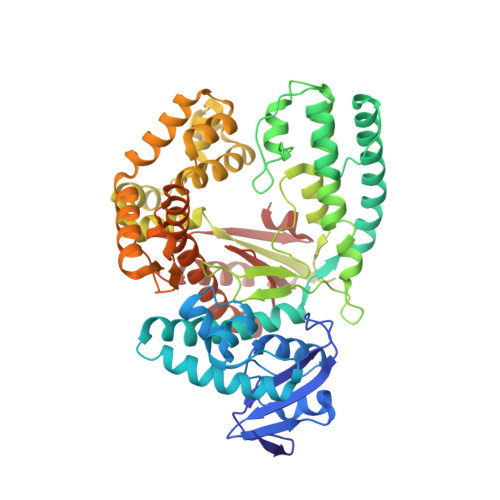
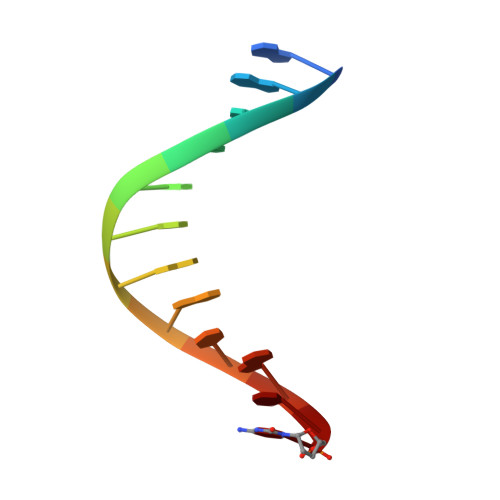
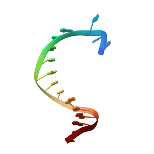
Interactions of non-polar and "Click-able" nucleotides in the confines of a DNA polymerase active site.

Obeid, S.Busskamp, H.Welte, W.Diederichs, K.Marx, A.

(2012) Chem Commun (Camb) 48: 8320-8322

  • DOI: https://doi.org/10.1039/c2cc34181f
  • Primary Citation of Related Structures:  
    4ELT, 4ELU

  • PubMed Abstract: 

    Modified nucleotides play a paramount role in many cutting-edge biomolecular techniques. The present structural study highlights the plasticity and flexibility of the active site of a DNA polymerase while incorporating non-polar "Click-able" nucleotide analogs and emphasizes new insights into rational design guidelines for modified nucleotides.


  • Organizational Affiliation

    Department of Chemistry and Konstanz Research School Chemical Biology, University of Konstanz, Universitätsstrasse 10, 78457 Konstanz, Germany.


Macromolecules

Find similar proteins by:  (by identity cutoff)  |  3D Structure
Entity ID: 1
MoleculeChains Sequence LengthOrganismDetailsImage
DNA polymerase I, thermostable540Thermus aquaticusMutation(s): 0 
Gene Names: polApol1
EC: 2.7.7.7
UniProt
Find proteins for P19821 (Thermus aquaticus)
Explore P19821 
Go to UniProtKB:  P19821
Entity Groups  
Sequence Clusters30% Identity50% Identity70% Identity90% Identity95% Identity100% Identity
UniProt GroupP19821
Sequence Annotations
Expand
  • Reference Sequence

Find similar nucleic acids by:  Sequence   |   3D Structure  

Entity ID: 2
MoleculeChains LengthOrganismImage
DNA (5'-D(*GP*AP*CP*CP*AP*CP*GP*GP*CP*GP*CP*(DOC))-3')12synthetic construct
Sequence Annotations
Expand
  • Reference Sequence

Find similar nucleic acids by:  Sequence   |   3D Structure  

Entity ID: 3
MoleculeChains LengthOrganismImage
DNA (5'-D(*AP*AP*AP*AP*GP*GP*CP*GP*CP*CP*GP*TP*GP*GP*TP*C)-3')16synthetic construct
Sequence Annotations
Expand
  • Reference Sequence
Small Molecules
Ligands 5 Unique
IDChains Name / Formula / InChI Key2D Diagram3D Interactions
0R5
Query on 0R5

Download Ideal Coordinates CCD File 
D [auth A]2'-deoxy-5-[(4-ethynylphenyl)ethynyl]uridine 5'-(tetrahydrogen triphosphate)
C19 H19 N2 O14 P3
NDHYKBWELLKQHS-GVDBMIGSSA-N
GOL
Query on GOL

Download Ideal Coordinates CCD File 
G [auth A]
I [auth A]
J [auth A]
L [auth A]
M [auth A]
G [auth A],
I [auth A],
J [auth A],
L [auth A],
M [auth A],
N [auth A],
P [auth A],
R [auth B],
S [auth C]
GLYCEROL
C3 H8 O3
PEDCQBHIVMGVHV-UHFFFAOYSA-N
EDO
Query on EDO

Download Ideal Coordinates CCD File 
Q [auth A]1,2-ETHANEDIOL
C2 H6 O2
LYCAIKOWRPUZTN-UHFFFAOYSA-N
ACT
Query on ACT

Download Ideal Coordinates CCD File 
H [auth A],
K [auth A],
O [auth A]
ACETATE ION
C2 H3 O2
QTBSBXVTEAMEQO-UHFFFAOYSA-M
MG
Query on MG

Download Ideal Coordinates CCD File 
E [auth A],
F [auth A],
T [auth C]
MAGNESIUM ION
Mg
JLVVSXFLKOJNIY-UHFFFAOYSA-N
Experimental Data & Validation

Experimental Data

  • Method: X-RAY DIFFRACTION
  • Resolution: 2.20 Å
  • R-Value Free: 0.209 
  • R-Value Work: 0.160 
  • R-Value Observed: 0.163 
  • Space Group: P 31 2 1
Unit Cell:
Length ( Å )Angle ( ˚ )
a = 108.86α = 90
b = 108.86β = 90
c = 91.004γ = 120
Software Package:
Software NamePurpose
XDSdata scaling
PHENIXmodel building
PHENIXrefinement
XDSdata reduction
PHENIXphasing

Structure Validation

View Full Validation Report


Ligand Structure Quality Assessment 


Entry History 

Deposition Data

Revision History  (Full details and data files)

  • Version 1.0: 2013-03-27
    Type: Initial release
  • Version 1.1: 2023-09-13
    Changes: Data collection, Database references, Derived calculations, Refinement description