4EHC

Crystal structure of the C-terminal domain of Rv0977 of Mycobacterium tuberculosis


Experimental Data Snapshot

  • Method: X-RAY DIFFRACTION
  • Resolution: 1.98 Å
  • R-Value Free: 0.213 
  • R-Value Work: 0.165 
  • R-Value Observed: 0.167 

wwPDB Validation   3D Report Full Report


This is version 1.1 of the entry. See complete history


Literature

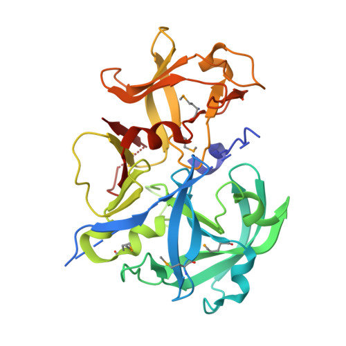
Crystal structure of a putative aspartic proteinase domain of the Mycobacterium tuberculosis cell surface antigen PE_PGRS16

Barathy, D.V.Suguna, K.

(2013) FEBS Open Bio 3: 256-262

  • DOI: https://doi.org/10.1016/j.fob.2013.05.004
  • Primary Citation of Related Structures:  
    4EHC

  • PubMed Abstract: 

    We report the crystal structure of the first prokaryotic aspartic proteinase-like domain identified in the genome of Mycobacterium tuberculosis. A search in the genomes of Mycobacterium species showed that the C-terminal domains of some of the PE family proteins contain two classic DT/SG motifs of aspartic proteinases with a low overall sequence similarity to HIV proteinase. The three-dimensional structure of one of them, Rv0977 (PE_PGRS16) of M. tuberculosis revealed the characteristic pepsin-fold and catalytic site architecture. However, the active site was completely blocked by the N-terminal His-tag. Surprisingly, the enzyme was found to be inactive even after the removal of the N-terminal His-tag. A comparison of the structure with pepsins showed significant differences in the critical substrate binding residues and in the flap tyrosine conformation that could contribute to the lack of proteolytic activity of Rv0977.


  • Organizational Affiliation

    Molecular Biophysics Unit, Indian Institute of Science, Bangalore, India.


Macromolecules
Find similar proteins by:  (by identity cutoff)  |  3D Structure
Entity ID: 1
MoleculeChains Sequence LengthOrganismDetailsImage
PE-PGRS FAMILY PROTEIN296Mycobacterium tuberculosisMutation(s): 0 
Gene Names: PE_PGRS16Rv0977
UniProt
Find proteins for Q79FU3 (Mycobacterium tuberculosis (strain ATCC 25618 / H37Rv))
Explore Q79FU3 
Go to UniProtKB:  Q79FU3
Entity Groups  
Sequence Clusters30% Identity50% Identity70% Identity90% Identity95% Identity100% Identity
UniProt GroupQ79FU3
Sequence Annotations
Expand
  • Reference Sequence
Small Molecules
Ligands 2 Unique
IDChains Name / Formula / InChI Key2D Diagram3D Interactions
ZN
Query on ZN

Download Ideal Coordinates CCD File 
B [auth A]ZINC ION
Zn
PTFCDOFLOPIGGS-UHFFFAOYSA-N
EDO
Query on EDO

Download Ideal Coordinates CCD File 
AA [auth A]
BA [auth A]
C [auth A]
CA [auth A]
D [auth A]
AA [auth A],
BA [auth A],
C [auth A],
CA [auth A],
D [auth A],
DA [auth A],
E [auth A],
EA [auth A],
F [auth A],
FA [auth A],
G [auth A],
GA [auth A],
H [auth A],
I [auth A],
J [auth A],
K [auth A],
L [auth A],
M [auth A],
N [auth A],
O [auth A],
P [auth A],
Q [auth A],
R [auth A],
S [auth A],
T [auth A],
U [auth A],
V [auth A],
W [auth A],
X [auth A],
Y [auth A],
Z [auth A]
1,2-ETHANEDIOL
C2 H6 O2
LYCAIKOWRPUZTN-UHFFFAOYSA-N
Modified Residues  1 Unique
IDChains TypeFormula2D DiagramParent
MSE
Query on MSE
A
L-PEPTIDE LINKINGC5 H11 N O2 SeMET
Experimental Data & Validation

Experimental Data

  • Method: X-RAY DIFFRACTION
  • Resolution: 1.98 Å
  • R-Value Free: 0.213 
  • R-Value Work: 0.165 
  • R-Value Observed: 0.167 
  • Space Group: P 21 21 21
Unit Cell:
Length ( Å )Angle ( ˚ )
a = 61.11α = 90
b = 66.16β = 90
c = 69.62γ = 90
Software Package:
Software NamePurpose
HKL-2000data collection
Auto-Rickshawphasing
REFMACrefinement
MOSFLMdata reduction
SCALAdata scaling

Structure Validation

View Full Validation Report


Entry History 

Deposition Data

Revision History  (Full details and data files)

  • Version 1.0: 2013-06-19
    Type: Initial release
  • Version 1.1: 2014-03-12
    Changes: Database references