4EF5

Crystal structure of STING CTD


Experimental Data Snapshot

  • Method: X-RAY DIFFRACTION
  • Resolution: 2.45 Å
  • R-Value Free: 0.288 
  • R-Value Work: 0.227 
  • R-Value Observed: 0.229 

wwPDB Validation   3D Report Full Report


This is version 1.4 of the entry. See complete history


Literature

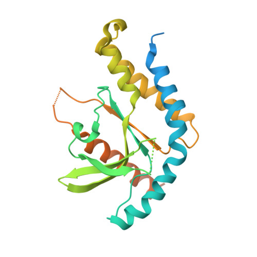
Structural analysis of the STING adaptor protein reveals a hydrophobic dimer interface and mode of cyclic di-GMP binding

Ouyang, S.Song, X.Wang, Y.Ru, H.Shaw, N.Jiang, Y.Niu, F.Zhu, Y.Qiu, W.Parvatiyar, K.Li, Y.Zhang, R.Cheng, G.Liu, Z.J.

(2012) Immunity 36: 1073-1086

  • DOI: https://doi.org/10.1016/j.immuni.2012.03.019
  • Primary Citation of Related Structures:  
    4EF4, 4EF5

  • PubMed Abstract: 

    STING is an essential signaling molecule for DNA and cyclic di-GMP (c-di-GMP)-mediated type I interferon (IFN) production via TANK-binding kinase 1 (TBK1) and interferon regulatory factor 3 (IRF3) pathway. It contains an N-terminal transmembrane region and a cytosolic C-terminal domain (CTD). Here, we describe crystal structures of STING CTD alone and complexed with c-di-GMP in a unique binding mode. The strictly conserved aa 153-173 region was shown to be cytosolic and participated in dimerization via hydrophobic interactions. The STING CTD functions as a dimer and the dimerization was independent of posttranslational modifications. Binding of c-di-GMP enhanced interaction of a shorter construct of STING CTD (residues 139-344) with TBK1. This suggests an extra TBK1 binding site, other than serine 358. This study provides a glimpse into the unique architecture of STING and sheds light on the mechanism of c-di-GMP-mediated TBK1 signaling.


  • Organizational Affiliation

    National Laboratory of Biomacromolecules, Institute of Biophysics, Chinese Academy of Sciences, Beijing, China.


Macromolecules
Find similar proteins by:  (by identity cutoff)  |  3D Structure
Entity ID: 1
MoleculeChains Sequence LengthOrganismDetailsImage
Transmembrane protein 173265Homo sapiensMutation(s): 0 
Gene Names: STING/MITA/ERIS/MPYS/TMEM173
UniProt & NIH Common Fund Data Resources
Find proteins for Q86WV6 (Homo sapiens)
Explore Q86WV6 
Go to UniProtKB:  Q86WV6
PHAROS:  Q86WV6
GTEx:  ENSG00000184584 
Entity Groups  
Sequence Clusters30% Identity50% Identity70% Identity90% Identity95% Identity100% Identity
UniProt GroupQ86WV6
Sequence Annotations
Expand
  • Reference Sequence
Experimental Data & Validation

Experimental Data

  • Method: X-RAY DIFFRACTION
  • Resolution: 2.45 Å
  • R-Value Free: 0.288 
  • R-Value Work: 0.227 
  • R-Value Observed: 0.229 
  • Space Group: C 2 2 21
Unit Cell:
Length ( Å )Angle ( ˚ )
a = 81.041α = 90
b = 90.356β = 90
c = 73.315γ = 90
Software Package:
Software NamePurpose
HKL-2000data collection
PHASESphasing
PHENIXrefinement
HKL-2000data reduction
HKL-2000data scaling

Structure Validation

View Full Validation Report


Entry History 

Deposition Data

Revision History  (Full details and data files)

  • Version 1.0: 2012-05-16
    Type: Initial release
  • Version 1.1: 2012-05-23
    Changes: Database references
  • Version 1.2: 2012-05-30
    Changes: Database references
  • Version 1.3: 2013-07-03
    Changes: Database references
  • Version 1.4: 2024-03-20
    Changes: Data collection, Database references