4EA0

Crystal structure of dehydrosqualene synthase (Crtm) from S. aureus complexed with diphosphate and quinuclidine BPH-651


Experimental Data Snapshot

  • Method: X-RAY DIFFRACTION
  • Resolution: 2.12 Å
  • R-Value Free: 0.278 
  • R-Value Work: 0.222 
  • R-Value Observed: 0.225 

wwPDB Validation   3D Report Full Report


Ligand Structure Quality Assessment 


This is version 1.2 of the entry. See complete history


Literature

Head-to-Head Prenyl Tranferases: Anti-Infective Drug Targets.

Lin, F.Y.Liu, Y.L.Li, K.Cao, R.Zhu, W.Axelson, J.Pang, R.Oldfield, E.

(2012) J Med Chem 55: 4367-4372

  • DOI: https://doi.org/10.1021/jm300208p
  • Primary Citation of Related Structures:  
    4E9U, 4E9Z, 4EA0, 4EA1, 4EA2

  • PubMed Abstract: 

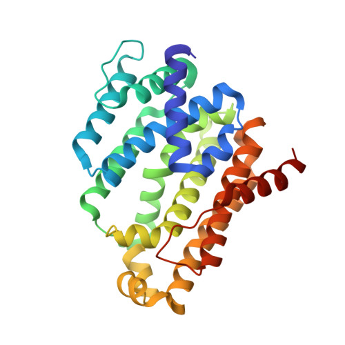
    We report X-ray crystallographic structures of three inhibitors bound to dehydrosqualene synthase from Staphylococcus aureus: 1 (BPH-651), 2 (WC-9), and 3 (SQ-109). Compound 2 binds to the S2 site with its -SCN group surrounded by four hydrogen bond donors. With 1, we report two structures: in both, the quinuclidine headgroup binds in the allylic (S1) site with the side chain in S2, but in the presence of PPi and Mg(2+), the quinuclidine's cationic center interacts with PPi and three Mg(2+), mimicking a transition state involved in diphosphate ionization. With 3, there are again two structures. In one, the geranyl side chain binds to either S1 or S2 and the adamantane headgroup binds to S1. In the second, the side chain binds to S2 while the headgroup binds to S1. These results provide structural clues for the mechanism and inhibition of the head-to-head prenyl transferases and should aid future drug design.


  • Organizational Affiliation

    Center for Biophysics and Computational Biology,University of Illinois at Urbana-Champaign, Urbana, Illinois 61801, USA.


Macromolecules
Find similar proteins by:  (by identity cutoff)  |  3D Structure
Entity ID: 1
MoleculeChains Sequence LengthOrganismDetailsImage
Dehydrosqualene synthase
A, B
287Staphylococcus aureusMutation(s): 0 
Gene Names: crtM
EC: 2.5.1.96
UniProt
Find proteins for A9JQL9 (Staphylococcus aureus)
Explore A9JQL9 
Go to UniProtKB:  A9JQL9
Entity Groups  
Sequence Clusters30% Identity50% Identity70% Identity90% Identity95% Identity100% Identity
UniProt GroupA9JQL9
Sequence Annotations
Expand
  • Reference Sequence
Small Molecules
Ligands 4 Unique
IDChains Name / Formula / InChI Key2D Diagram3D Interactions
651
Query on 651

Download Ideal Coordinates CCD File 
H [auth A],
M [auth B]
(3R)-3-biphenyl-4-yl-1-azabicyclo[2.2.2]octan-3-ol
C19 H21 N O
WPCQYFUQHBLGAX-IBGZPJMESA-N
POP
Query on POP

Download Ideal Coordinates CCD File 
F [auth A],
L [auth B]
PYROPHOSPHATE 2-
H2 O7 P2
XPPKVPWEQAFLFU-UHFFFAOYSA-L
TLA
Query on TLA

Download Ideal Coordinates CCD File 
G [auth A]L(+)-TARTARIC ACID
C4 H6 O6
FEWJPZIEWOKRBE-JCYAYHJZSA-N
MG
Query on MG

Download Ideal Coordinates CCD File 
C [auth A]
D [auth A]
E [auth A]
I [auth B]
J [auth B]
C [auth A],
D [auth A],
E [auth A],
I [auth B],
J [auth B],
K [auth B]
MAGNESIUM ION
Mg
JLVVSXFLKOJNIY-UHFFFAOYSA-N
Experimental Data & Validation

Experimental Data

  • Method: X-RAY DIFFRACTION
  • Resolution: 2.12 Å
  • R-Value Free: 0.278 
  • R-Value Work: 0.222 
  • R-Value Observed: 0.225 
  • Space Group: P 31 2 1
Unit Cell:
Length ( Å )Angle ( ˚ )
a = 80.324α = 90
b = 80.324β = 90
c = 180.822γ = 120
Software Package:
Software NamePurpose
PHASESphasing
REFMACrefinement
HKL-2000data reduction
HKL-2000data scaling

Structure Validation

View Full Validation Report


Ligand Structure Quality Assessment 


Entry History 

Deposition Data

Revision History  (Full details and data files)

  • Version 1.0: 2012-04-25
    Type: Initial release
  • Version 1.1: 2012-05-16
    Changes: Database references
  • Version 1.2: 2024-02-28
    Changes: Data collection, Database references, Derived calculations