4DXH

Horse liver alcohol dehydrogenase complexed with NAD+ and 2,2,2-trifluoroethanol


Experimental Data Snapshot

  • Method: X-RAY DIFFRACTION
  • Resolution: 1.12 Å
  • R-Value Free: 0.147 
  • R-Value Work: 0.127 
  • R-Value Observed: 0.127 

wwPDB Validation   3D Report Full Report


Ligand Structure Quality Assessment 


This is version 1.6 of the entry. See complete history


Literature

Atomic-Resolution Structures of Horse Liver Alcohol Dehydrogenase with NAD(+) and Fluoroalcohols Define Strained Michaelis Complexes.

Plapp, B.V.Ramaswamy, S.

(2012) Biochemistry 51: 4035-4048

  • DOI: https://doi.org/10.1021/bi300378n
  • Primary Citation of Related Structures:  
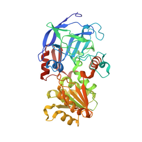
    4DWV, 4DXH

  • PubMed Abstract: 

    Structures of horse liver alcohol dehydrogenase complexed with NAD(+) and unreactive substrate analogues, 2,2,2-trifluoroethanol or 2,3,4,5,6-pentafluorobenzyl alcohol, were determined at 100 K at 1.12 or 1.14 Å resolution, providing estimates of atomic positions with overall errors of ~0.02 Å, the geometry of ligand binding, descriptions of alternative conformations of amino acid residues and waters, and evidence of a strained nicotinamide ring. The four independent subunits from the two homodimeric structures differ only slightly in the peptide backbone conformation. Alternative conformations for amino acid side chains were identified for 50 of the 748 residues in each complex, and Leu-57 and Leu-116 adopt different conformations to accommodate the different alcohols at the active site. Each fluoroalcohol occupies one position, and the fluorines of the alcohols are well-resolved. These structures closely resemble the expected Michaelis complexes with the pro-R hydrogens of the methylene carbons of the alcohols directed toward the re face of C4N of the nicotinamide rings with a C-C distance of 3.40 Å. The oxygens of the alcohols are ligated to the catalytic zinc at a distance expected for a zinc alkoxide (1.96 Å) and participate in a low-barrier hydrogen bond (2.52 Å) with the hydroxyl group of Ser-48 in a proton relay system. As determined by X-ray refinement with no restraints on bond distances and planarity, the nicotinamide rings in the two complexes are slightly puckered (quasi-boat conformation, with torsion angles of 5.9° for C4N and 4.8° for N1N relative to the plane of the other atoms) and have bond distances that are somewhat different compared to those found for NAD(P)(+). It appears that the nicotinamide ring is strained toward the transition state on the path to alcohol oxidation.


  • Organizational Affiliation

    Department of Biochemistry, The University of Iowa, Iowa City, IA 52242, USA. bv-plapp@uiowa.edu


Macromolecules
Find similar proteins by:  (by identity cutoff)  |  3D Structure
Entity ID: 1
MoleculeChains Sequence LengthOrganismDetailsImage
Alcohol dehydrogenase E chain
A, B
374Equus caballusMutation(s): 0 
EC: 1.1.1.1
UniProt
Find proteins for P00327 (Equus caballus)
Explore P00327 
Go to UniProtKB:  P00327
Entity Groups  
Sequence Clusters30% Identity50% Identity70% Identity90% Identity95% Identity100% Identity
UniProt GroupP00327
Sequence Annotations
Expand
  • Reference Sequence
Small Molecules
Ligands 4 Unique
IDChains Name / Formula / InChI Key2D Diagram3D Interactions
NAJ
Query on NAJ

Download Ideal Coordinates CCD File 
E [auth A],
L [auth B]
NICOTINAMIDE-ADENINE-DINUCLEOTIDE (ACIDIC FORM)
C21 H27 N7 O14 P2
BAWFJGJZGIEFAR-NNYOXOHSSA-N
MRD
Query on MRD

Download Ideal Coordinates CCD File 
G [auth A],
H [auth A],
I [auth A],
N [auth B]
(4R)-2-METHYLPENTANE-2,4-DIOL
C6 H14 O2
SVTBMSDMJJWYQN-RXMQYKEDSA-N
ETF
Query on ETF

Download Ideal Coordinates CCD File 
F [auth A],
M [auth B]
TRIFLUOROETHANOL
C2 H3 F3 O
RHQDFWAXVIIEBN-UHFFFAOYSA-N
ZN
Query on ZN

Download Ideal Coordinates CCD File 
C [auth A],
D [auth A],
J [auth B],
K [auth B]
ZINC ION
Zn
PTFCDOFLOPIGGS-UHFFFAOYSA-N
Binding Affinity Annotations 
IDSourceBinding Affinity
ETF Binding MOAD:  4DXH Ki: 8400 (nM) from 1 assay(s)
Experimental Data & Validation

Experimental Data

Unit Cell:
Length ( Å )Angle ( ˚ )
a = 44.25α = 91.91
b = 51.16β = 103.02
c = 92.53γ = 109.9
Software Package:
Software NamePurpose
REFMACrefinement
PDB_EXTRACTdata extraction
ADSCdata collection
d*TREKdata reduction
d*TREKdata scaling
AMoREphasing

Structure Validation

View Full Validation Report


Ligand Structure Quality Assessment 


Entry History 

Deposition Data

Revision History  (Full details and data files)

  • Version 1.0: 2012-04-11
    Type: Initial release
  • Version 1.1: 2012-05-02
    Changes: Structure summary
  • Version 1.2: 2012-05-16
    Changes: Database references
  • Version 1.3: 2012-06-27
    Changes: Database references
  • Version 1.4: 2022-04-27
    Changes: Database references, Derived calculations
  • Version 1.5: 2022-10-12
    Changes: Database references
  • Version 1.6: 2023-09-20
    Changes: Data collection, Refinement description