4DUH

Crystal structure of 24 kDa domain of E. coli DNA gyrase B in complex with small molecule inhibitor


Experimental Data Snapshot

  • Method: X-RAY DIFFRACTION
  • Resolution: 1.50 Å
  • R-Value Free: 0.226 
  • R-Value Work: 0.192 
  • R-Value Observed: 0.193 

wwPDB Validation   3D Report Full Report


Ligand Structure Quality Assessment 


This is version 1.2 of the entry. See complete history


Literature

Structure-based discovery of substituted 4,5'-bithiazoles as novel DNA gyrase inhibitors.

Brvar, M.Perdih, A.Renko, M.Anderluh, G.Turk, D.Solmajer, T.

(2012) J Med Chem 55: 6413-6426

  • DOI: https://doi.org/10.1021/jm300395d
  • Primary Citation of Related Structures:  
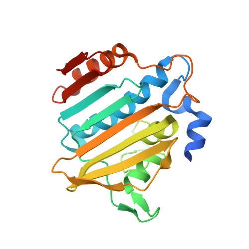
    4DUH

  • PubMed Abstract: 

    Bacterial DNA gyrase is a well-established and validated target for the development of novel antibacterials. Starting from the available structural information about the binding of the natural product inhibitor, clorobiocin, we identified a novel series of 4'-methyl-N(2)-phenyl-[4,5'-bithiazole]-2,2'-diamine inhibitors of gyrase B with a low micromolar inhibitory activity by implementing a two-step structure-based design procedure. This novel class of DNA gyrase inhibitors was extensively investigated by various techniques (differential scanning fluorimetry, surface plasmon resonance, and microscale thermophoresis). The binding mode of the potent inhibitor 18 was revealed by X-ray crystallography, confirming our initial in silico binding model. Furthermore, the high resolution of the complex structure allowed for the placement of the Gly97-Ser108 flexible loop, thus revealing its role in binding of this class of compounds. The crystal structure of the complex protein G24 and inhibitor 18 provides valuable information for further optimization of this novel class of DNA gyrase B inhibitors.


  • Organizational Affiliation

    National Institute of Chemistry, Laboratory for Biocomputing and Bioinformatics, 1001 Ljubljana, Slovenia.


Macromolecules
Find similar proteins by:  (by identity cutoff)  |  3D Structure
Entity ID: 1
MoleculeChains Sequence LengthOrganismDetailsImage
DNA gyrase subunit B
A, B
220Escherichia coli K-12Mutation(s): 0 
Gene Names: gyrBacrBhimBhisUnalCparApcbAb3699JW5625
EC: 5.99.1.3
UniProt
Find proteins for P0AES6 (Escherichia coli (strain K12))
Explore P0AES6 
Go to UniProtKB:  P0AES6
Entity Groups  
Sequence Clusters30% Identity50% Identity70% Identity90% Identity95% Identity100% Identity
UniProt GroupP0AES6
Sequence Annotations
Expand
  • Reference Sequence
Small Molecules
Ligands 1 Unique
IDChains Name / Formula / InChI Key2D Diagram3D Interactions
RLI
Query on RLI

Download Ideal Coordinates CCD File 
C [auth A],
D [auth B]
4-{[4'-methyl-2'-(propanoylamino)-4,5'-bi-1,3-thiazol-2-yl]amino}benzoic acid
C17 H16 N4 O3 S2
JOAXMWDTALNZFE-UHFFFAOYSA-N
Binding Affinity Annotations 
IDSourceBinding Affinity
RLI PDBBind:  4DUH Kd: 6600 (nM) from 1 assay(s)
Binding MOAD:  4DUH Kd: 6600 (nM) from 1 assay(s)
Experimental Data & Validation

Experimental Data

  • Method: X-RAY DIFFRACTION
  • Resolution: 1.50 Å
  • R-Value Free: 0.226 
  • R-Value Work: 0.192 
  • R-Value Observed: 0.193 
  • Space Group: P 1 21 1
Unit Cell:
Length ( Å )Angle ( ˚ )
a = 53.236α = 90
b = 51.122β = 95.68
c = 79.13γ = 90
Software Package:
Software NamePurpose
DENZOdata reduction
SCALEPACKdata scaling
REFMACrefinement
PDB_EXTRACTdata extraction
MxCuBEdata collection
PHASERphasing

Structure Validation

View Full Validation Report


Ligand Structure Quality Assessment 


Entry History 

Deposition Data

Revision History  (Full details and data files)

  • Version 1.0: 2012-08-01
    Type: Initial release
  • Version 1.1: 2012-08-08
    Changes: Database references
  • Version 1.2: 2023-09-13
    Changes: Data collection, Database references, Derived calculations, Refinement description