Structure of a yeast Dyn2-Nup159 complex and molecular basis for dynein light chain-nuclear pore interaction.
Romes, E.M., Tripathy, A., Slep, K.C.(2012) J Biol Chem 287: 15862-15873
- PubMed: 22411995
- DOI: https://doi.org/10.1074/jbc.M111.336172
- Primary Citation of Related Structures:
4DS1 - PubMed Abstract:
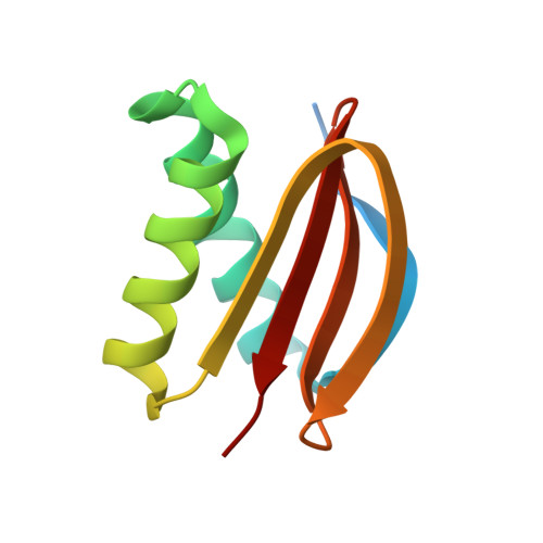
The nuclear pore complex gates nucleocytoplasmic transport through a massive, eight-fold symmetric channel capped by a nucleoplasmic basket and structurally unique, cytoplasmic fibrils whose tentacles bind and regulate asymmetric traffic. The conserved Nup82 complex, composed of Nsp1, Nup82, and Nup159, forms the unique cytoplasmic fibrils that regulate mRNA nuclear export. Although the nuclear pore complex plays a fundamental, conserved role in nuclear trafficking, structural information about the cytoplasmic fibrils is limited. Here, we investigate the structural and biochemical interactions between Saccharomyces cerevisiae Nup159 and the nucleoporin, Dyn2. We find that Dyn2 is predominantly a homodimer and binds arrayed sites on Nup159, promoting the Nup159 parallel homodimerization. We present the first structure of Dyn2, determined at 1.85 Å resolution, complexed with a Nup159 target peptide. Dyn2 resembles homologous metazoan dynein light chains, forming homodimeric composite substrate binding sites that engage two independent 10-residue target motifs, imparting a β-strand structure to each peptide via antiparallel extension of the Dyn2 core β-sandwich. Dyn2 recognizes a highly conserved QT motif while allowing sequence plasticity in the flanking residues of the peptide. Isothermal titration calorimetric analysis of the comparative binding of Dyn2 to two Nup159 target sites shows similar affinities (18 and 13 μM), but divergent thermal binding modes. Dyn2 homodimers are arrayed in the crystal lattice, likely mimicking the arrayed architecture of Dyn2 on the Nup159 multivalent binding sites. Crystallographic interdimer interactions potentially reflect a cooperative basis for Dyn2-Nup159 complex formation. Our data highlight the determinants that mediate oligomerization of the Nup82 complex and promote a directed, elongated cytoplasmic fibril architecture.
Organizational Affiliation:
Department of Biochemistry and Biophysics, University of North Carolina, Chapel Hill, North Carolina 27599, USA.