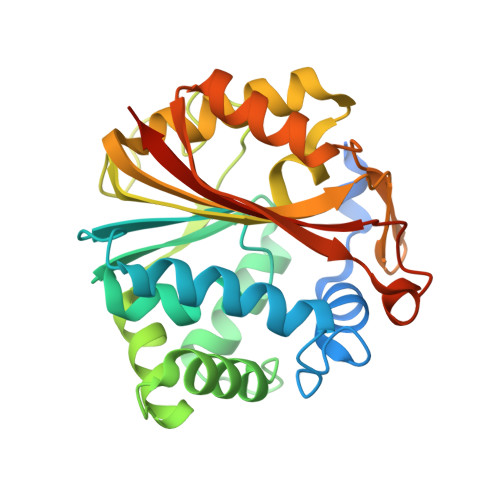
4DM3

Crystal structure of human PNMT in complex adohcy, resorcinol and imidazole


Experimental Data Snapshot

  • Method: X-RAY DIFFRACTION
  • Resolution: 2.40 Å
  • R-Value Free: 0.275 
  • R-Value Work: 0.220 
  • R-Value Observed: 0.223 

wwPDB Validation   3D Report Full Report


Ligand Structure Quality Assessment 


This is version 1.2 of the entry. See complete history

Re-refinement Note

This entry reflects an alternative modeling of the original data in: 3KPY


Literature

Missing fragments: detecting cooperative binding in fragment-based drug design.

Nair, P.C.Malde, A.K.Drinkwater, N.Mark, A.E.

(2012) ACS Med Chem Lett 3: 322-326

  • DOI: https://doi.org/10.1021/ml300015u
  • Primary Citation of Related Structures:  
    4DM3

  • PubMed Abstract: 

    The aim of fragment-based drug design (FBDD) is to identify molecular fragments that bind to alternate subsites within a given binding pocket leading to cooperative binding when linked. In this study, the binding of fragments to human phenylethanolamine N-methyltransferase is used to illustrate how (a) current protocols may fail to detect fragments that bind cooperatively, (b) theoretical approaches can be used to validate potential hits, and (c) apparent false positives obtained when screening against cocktails of fragments may in fact indicate promising leads.


  • Organizational Affiliation

    School of Chemistry and Molecular Biosciences (SCMB) and Institute for Molecular Bioscience (IMB), The University of Queensland (UQ) , St Lucia Campus, Brisbane, QLD 4072, Australia.


Macromolecules
Find similar proteins by:  (by identity cutoff)  |  3D Structure
Entity ID: 1
MoleculeChains Sequence LengthOrganismDetailsImage
Phenylethanolamine N-methyltransferase
A, B
289Homo sapiensMutation(s): 0 
Gene Names: PNMTPENT
EC: 2.1.1.28
UniProt & NIH Common Fund Data Resources
Find proteins for P11086 (Homo sapiens)
Explore P11086 
Go to UniProtKB:  P11086
PHAROS:  P11086
GTEx:  ENSG00000141744 
Entity Groups  
Sequence Clusters30% Identity50% Identity70% Identity90% Identity95% Identity100% Identity
UniProt GroupP11086
Sequence Annotations
Expand
  • Reference Sequence
Experimental Data & Validation

Experimental Data

  • Method: X-RAY DIFFRACTION
  • Resolution: 2.40 Å
  • R-Value Free: 0.275 
  • R-Value Work: 0.220 
  • R-Value Observed: 0.223 
  • Space Group: P 43 21 2
Unit Cell:
Length ( Å )Angle ( ˚ )
a = 93.908α = 90
b = 93.908β = 90
c = 188.615γ = 90
Software Package:
Software NamePurpose
PHENIXrefinement

Structure Validation

View Full Validation Report


Ligand Structure Quality Assessment 


Entry History 

Deposition Data

Revision History  (Full details and data files)

  • Version 1.0: 2012-04-25
    Type: Initial release
  • Version 1.1: 2014-09-17
    Changes: Database references
  • Version 1.2: 2018-01-24
    Changes: Advisory, Structure summary