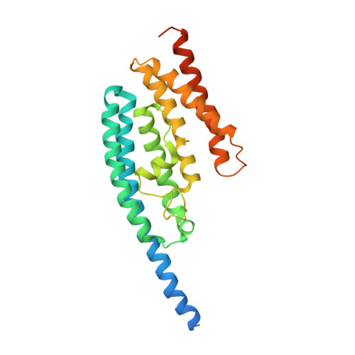
Low-resolution structure of Drosophila translin
Kumar, V., Gupta, G.D.(2012) FEBS Open Bio 2: 37-46
- PubMed: 23650579
- DOI: https://doi.org/10.1016/j.fob.2012.03.001
- Primary Citation of Related Structures:
4DG7 - PubMed Abstract:
Crystals of native Drosophila melanogaster translin diffracted to 7 Å resolution. Reductive methylation of the protein improved crystal quality. The native and methylated proteins showed similar profiles in size-exclusion chromatography analyses but the methylated protein displayed reduced DNA-binding activity. Crystals of the methylated protein diffracted to 4.2 Å resolution at BM14 of the ESRF synchrotron. Crystals with 49% solvent content belonged to monoclinic space group P21 with eight protomers in the asymmetric unit. Only 2% of low-resolution structures with similar low percentage solvent content were found in the PDB. The crystal structure, solved by molecular replacement method, refined to R work (R free) of 0.24 (0.29) with excellent stereochemistry. The crystal structure clearly shows that drosophila protein exists as an octamer, and not as a decamer as expected from gel-filtration elution profiles. The similar octameric quaternary fold in translin orthologs and in translin-TRAX complexes suggests an up-down dimer as the basic structural subunit of translin-like proteins. The drosophila oligomer displays asymmetric assembly and increased radius of gyration that accounts for the observed differences between the elution profiles of human and drosophila proteins on gel-filtration columns. This study demonstrates clearly that low-resolution X-ray structure can be useful in understanding complex biological oligomers.
Organizational Affiliation:
High Pressure & Synchrotron Radiation Physics Division, Bhabha Atomic Research Centre, Mumbai 400085, India.