4DFG

Crystal Structure of Wild-type HIV-1 Protease with Cyclopentyltetrahydro- furanyl Urethanes as P2-ligand, GRL-0249A


Experimental Data Snapshot

  • Method: X-RAY DIFFRACTION
  • Resolution: 1.23 Å
  • R-Value Free: 0.180 
  • R-Value Work: 0.144 
  • R-Value Observed: 0.146 

wwPDB Validation   3D Report Full Report


Ligand Structure Quality Assessment 


This is version 1.1 of the entry. See complete history


Literature

Substituent effects on P2-cyclopentyltetrahydrofuranyl urethanes: Design, synthesis, and X-ray studies of potent HIV-1 protease inhibitors.

Ghosh, A.K.Chapsal, B.D.Steffey, M.Agniswamy, J.Wang, Y.F.Amano, M.Weber, I.T.Mitsuya, H.

(2012) Bioorg Med Chem Lett 22: 2308-2311

  • DOI: https://doi.org/10.1016/j.bmcl.2012.01.061
  • Primary Citation of Related Structures:  
    4DFG

  • PubMed Abstract: 

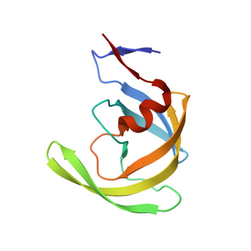
    The design, synthesis, and biological evaluation of novel C3-substituted cyclopentyltetrahydrofuranyl (Cp-THF)-derived HIV-1 protease inhibitors are described. Various C3-functional groups on the Cp-THF ligand were investigated in order to maximize the ligand-binding site interactions in the flap region of the protease. Inhibitors 3c and 3d have displayed the most potent enzyme inhibitory and antiviral activity. Both inhibitors have maintained impressive activity against a panel of multidrug resistant HIV-1 variants. A high-resolution X-ray crystal structure of 3c-bound HIV-1 protease revealed a number of important molecular insights into the ligand-binding site interactions.


  • Organizational Affiliation

    Department of Chemistry, Purdue University, West Lafayette, IN 47907, USA. akghosh@purdue.edu


Macromolecules
Find similar proteins by:  (by identity cutoff)  |  3D Structure
Entity ID: 1
MoleculeChains Sequence LengthOrganismDetailsImage
Protease
A, B
99Human immunodeficiency virus type 1 (BRU ISOLATE)Mutation(s): 5 
Gene Names: gag-pol
EC: 3.4.23.16
UniProt
Find proteins for P03367 (Human immunodeficiency virus type 1 group M subtype B (isolate BRU/LAI))
Explore P03367 
Go to UniProtKB:  P03367
Entity Groups  
Sequence Clusters30% Identity50% Identity70% Identity90% Identity95% Identity100% Identity
UniProt GroupP03367
Sequence Annotations
Expand
  • Reference Sequence
Small Molecules
Ligands 3 Unique
IDChains Name / Formula / InChI Key2D Diagram3D Interactions
0JV
Query on 0JV

Download Ideal Coordinates CCD File 
F [auth B]methyl N-[(3S,3aR,5R,6aR)-5-[[(2S,3R)-4-[(4-methoxyphenyl)sulfonyl-(2-methylpropyl)amino]-3-oxidanyl-1-phenyl-butan-2-yl]carbamoyloxy]-3,3a,4,5,6,6a-hexahydro-2H-cyclopenta[b]furan-3-yl]carbamate
C31 H43 N3 O9 S
XXQYXAWGNBWMGJ-YZGPKJBYSA-N
CL
Query on CL

Download Ideal Coordinates CCD File 
D [auth A],
E [auth A],
H [auth B],
I [auth B]
CHLORIDE ION
Cl
VEXZGXHMUGYJMC-UHFFFAOYSA-M
NA
Query on NA

Download Ideal Coordinates CCD File 
C [auth A],
G [auth B]
SODIUM ION
Na
FKNQFGJONOIPTF-UHFFFAOYSA-N
Binding Affinity Annotations 
IDSourceBinding Affinity
0JV Binding MOAD:  4DFG Ki: 1.80e-3 (nM) from 1 assay(s)
PDBBind:  4DFG Ki: 1.80e-3 (nM) from 1 assay(s)
BindingDB:  4DFG Ki: 1.8 (nM) from 1 assay(s)
Experimental Data & Validation

Experimental Data

  • Method: X-RAY DIFFRACTION
  • Resolution: 1.23 Å
  • R-Value Free: 0.180 
  • R-Value Work: 0.144 
  • R-Value Observed: 0.146 
  • Space Group: P 21 21 2
Unit Cell:
Length ( Å )Angle ( ˚ )
a = 58.58α = 90
b = 85.93β = 90
c = 45.99γ = 90
Software Package:
Software NamePurpose
HKL-2000data collection
PHASESphasing
SHELXL-97refinement
HKL-2000data reduction
HKL-2000data scaling

Structure Validation

View Full Validation Report


Ligand Structure Quality Assessment 


Entry History 

Deposition Data

Revision History  (Full details and data files)

  • Version 1.0: 2012-03-21
    Type: Initial release
  • Version 1.1: 2023-09-13
    Changes: Data collection, Database references, Derived calculations, Refinement description