4D7W

Crystal structure of sortase C1 (SrtC1) from Streptococcus agalactiae


Experimental Data Snapshot

  • Method: X-RAY DIFFRACTION
  • Resolution: 1.95 Å
  • R-Value Free: 0.223 
  • R-Value Work: 0.180 
  • R-Value Observed: 0.182 

wwPDB Validation   3D Report Full Report


This is version 2.1 of the entry. See complete history


Literature

Noncanonical Sortase-Mediated Assembly of Pilus Type 2B in Group B Streptococcus.

Lazzarin, M.Cozzi, R.Malito, E.Martinelli, M.D'Onofrio, M.Maione, D.Margarit, I.Rinaudo, C.D.

(2015) FASEB J 29: 4629

  • DOI: https://doi.org/10.1096/fj.15-272500
  • Primary Citation of Related Structures:  
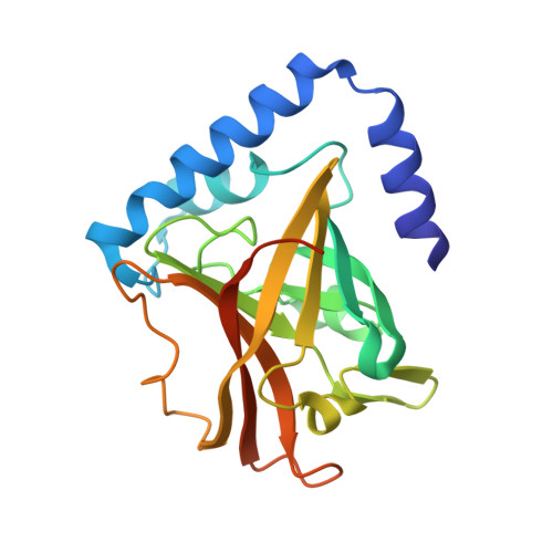
    4D7W

  • PubMed Abstract: 

    Group B Streptococcus (GBS) expresses 3 structurally distinct pilus types (1, 2a, and 2b) identified as important virulence factors and vaccine targets. These pili are heterotrimeric polymers, covalently assembled on the cell wall by sortase (Srt) enzymes. We investigated the pilus-2b biogenesis mechanism by using a multidisciplinary approach integrating genetic, biochemical, and structural studies to dissect the role of the 2 pilus-2b-associated Srts. We show that only 1 sortase (SrtC1-2b) is responsible for pilus protein polymerization, whereas the second one (Srt2-2b) does not act as a pilin polymerase, but similarly to the housekeeping class A Srt (SrtA), it is involved in cell-wall pilus anchoring by targeting the minor ancillary subunit. Based on its function and sequence features, Srt2-2b does not belong to class C Srts (SrtCs), nor is it a canonical member of any other known family of Srts. We also report the crystal structure of SrtC1-2b at 1.9 Å resolution. The overall fold resembles the typical structure of SrtCs except for the N-terminal lid region that appears in an open conformation displaced from the active site. Our findings reveal that GBS pilus type 2b biogenesis differs significantly from the current model of pilus assembly in gram-positive pathogens.


  • Organizational Affiliation

    *Novartis Vaccines and Diagnostics, GlaxoSmithKline, Siena, Italy; and Nuclear Magnetic Resonance Laboratory, Department of Biotechnology, University of Verona, Verona, Italy.


Macromolecules
Find similar proteins by:  (by identity cutoff)  |  3D Structure
Entity ID: 1
MoleculeChains Sequence LengthOrganismDetailsImage
SORTASE FAMILY PROTEIN208Streptococcus agalactiae COH1Mutation(s): 0 
UniProt
Find proteins for A0A0M3KKU7 (Streptococcus agalactiae serotype III (strain COH1))
Explore A0A0M3KKU7 
Go to UniProtKB:  A0A0M3KKU7
Entity Groups  
Sequence Clusters30% Identity50% Identity70% Identity90% Identity95% Identity100% Identity
UniProt GroupA0A0M3KKU7
Sequence Annotations
Expand
  • Reference Sequence
Small Molecules
Ligands 1 Unique
IDChains Name / Formula / InChI Key2D Diagram3D Interactions
EDO
Query on EDO

Download Ideal Coordinates CCD File 
B [auth A]1,2-ETHANEDIOL
C2 H6 O2
LYCAIKOWRPUZTN-UHFFFAOYSA-N
Experimental Data & Validation

Experimental Data

  • Method: X-RAY DIFFRACTION
  • Resolution: 1.95 Å
  • R-Value Free: 0.223 
  • R-Value Work: 0.180 
  • R-Value Observed: 0.182 
  • Space Group: P 21 21 2
Unit Cell:
Length ( Å )Angle ( ˚ )
a = 37.56α = 90
b = 124.76β = 90
c = 45.92γ = 90
Software Package:
Software NamePurpose
PHENIXrefinement
XDSdata reduction
XSCALEdata scaling
MrBUMPphasing

Structure Validation

View Full Validation Report


Entry History 

Deposition Data

Revision History  (Full details and data files)

  • Version 1.0: 2015-08-19
    Type: Initial release
  • Version 1.1: 2015-11-18
    Changes: Database references
  • Version 2.0: 2017-09-27
    Changes: Atomic model, Data collection
  • Version 2.1: 2023-12-20
    Changes: Data collection, Database references, Derived calculations, Other, Refinement description