Structure and Sialyllactose Binding of the Carboxy-Terminal Head Domain of the Fibre from a Siadenovirus, Turkey Adenovirus 3.
Singh, A.K., Berbis, M.A., Ballmann, M.Z., Kilcoyne, M., Menendez, M., Nguyen, T.H., Joshi, L., Canada, F.J., Jimenez-Barbero, J., Benko, M., Harrach, B., van Raaij, M.J.(2015) PLoS One 10: e0139339-e0139339
- PubMed: 26418008
- DOI: https://doi.org/10.1371/journal.pone.0139339
- Primary Citation of Related Structures:
3ZPE, 3ZPF, 4CW8, 4D62, 4D63 - PubMed Abstract:
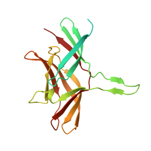
The virulent form of turkey adenovirus 3 (TAdV-3), also known as turkey hemorrhagic enteritis virus (THEV), is an economically important poultry pathogen, while the avirulent form is used as a vaccine. TAdV-3 belongs to the genus Siadenovirus. The carboxy-terminal region of its fibre does not have significant sequence similarity to any other adenovirus fibre heads of known structure. Two amino acid sequence differences between virulent and avirulent TAdV-3 map on the fibre head: where virulent TAdV-3 contains Ile354 and Thr376, avirulent TAdV-3 contains Met354 and Met376. We determined the crystal structures of the trimeric virulent and avirulent TAdV-3 fibre head domains at 2.2 Å resolution. Each monomer contains a beta-sandwich, which, surprisingly, resembles reovirus fibre head more than other adenovirus fibres, although the ABCJ-GHID topology is conserved in all. A beta-hairpin insertion in the C-strand of each trimer subunit embraces its neighbouring monomer. The avirulent and virulent TAdV-3 fibre heads are identical apart from the exact orientation of the beta-hairpin insertion. In vitro, sialyllactose was identified as a ligand by glycan microarray analysis, nuclear magnetic resonance spectroscopy, and crystallography. Its dissociation constant was measured to be in the mM range by isothermal titration calorimetry. The ligand binds to the side of the fibre head, involving amino acids Glu392, Thr419, Val420, Lys421, Asn422, and Gly423 binding to the sialic acid group. It binds slightly more strongly to the avirulent form. We propose that, in vivo, the TAdV-3 fibre may bind a sialic acid-containing cell surface component.
Organizational Affiliation:
Departamento de Estructura de Macromoléculas, Centro Nacional de Biotecnología (CNB-CSIC), Madrid, Spain.