4D1E

THE CRYSTAL STRUCTURE OF HUMAN MUSCLE ALPHA-ACTININ-2


Experimental Data Snapshot

  • Method: X-RAY DIFFRACTION
  • Resolution: 3.50 Å
  • R-Value Free: 0.258 
  • R-Value Work: 0.205 
  • R-Value Observed: 0.207 

wwPDB Validation   3D Report Full Report


This is version 1.2 of the entry. See complete history


Literature

The Structure and Regulation of Human Muscle Alpha-Actinin

Ribeiro Jr, E.A.Pinotsis, N.Ghisleni, A.Salmazo, A.Konarev, P.V.Kostan, J.Sjoeblom, B.Schreiner, C.Polyansky, A.A.Gkougkoulia, E.Holt, M.R.Aachmann, F.L.Zagrovic, B.Bordignon, E.Pirker, K.F.Svergun, D.I.Gautel, M.Djinovic-Carugo, K.

(2014) Cell 159: 1447

  • DOI: https://doi.org/10.1016/j.cell.2014.10.056
  • Primary Citation of Related Structures:  
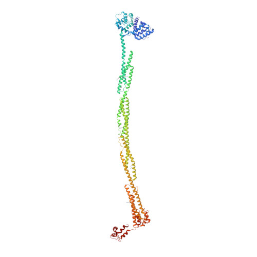
    4D1E

  • PubMed Abstract: 

    The spectrin superfamily of proteins plays key roles in assembling the actin cytoskeleton in various cell types, crosslinks actin filaments, and acts as scaffolds for the assembly of large protein complexes involved in structural integrity and mechanosensation, as well as cell signaling. α-actinins in particular are the major actin crosslinkers in muscle Z-disks, focal adhesions, and actin stress fibers. We report a complete high-resolution structure of the 200 kDa α-actinin-2 dimer from striated muscle and explore its functional implications on the biochemical and cellular level. The structure provides insight into the phosphoinositide-based mechanism controlling its interaction with sarcomeric proteins such as titin, lays a foundation for studying the impact of pathogenic mutations at molecular resolution, and is likely to be broadly relevant for the regulation of spectrin-like proteins.


  • Organizational Affiliation

    Department of Structural and Computational Biology, Max F. Perutz Laboratories, University of Vienna, Campus Vienna Biocenter 5, 1030 Vienna, Austria.


Macromolecules
Find similar proteins by:  (by identity cutoff)  |  3D Structure
Entity ID: 1
MoleculeChains Sequence LengthOrganismDetailsImage
ALPHA-ACTININ-2876Homo sapiensMutation(s): 2 
UniProt & NIH Common Fund Data Resources
Find proteins for P35609 (Homo sapiens)
Explore P35609 
Go to UniProtKB:  P35609
PHAROS:  P35609
GTEx:  ENSG00000077522 
Entity Groups  
Sequence Clusters30% Identity50% Identity70% Identity90% Identity95% Identity100% Identity
UniProt GroupP35609
Sequence Annotations
Expand
  • Reference Sequence
Small Molecules
Modified Residues  1 Unique
IDChains TypeFormula2D DiagramParent
MLZ
Query on MLZ
A
L-PEPTIDE LINKINGC7 H16 N2 O2LYS
Experimental Data & Validation

Experimental Data

  • Method: X-RAY DIFFRACTION
  • Resolution: 3.50 Å
  • R-Value Free: 0.258 
  • R-Value Work: 0.205 
  • R-Value Observed: 0.207 
  • Space Group: P 2 21 21
Unit Cell:
Length ( Å )Angle ( ˚ )
a = 73.334α = 90
b = 102.613β = 90
c = 182.977γ = 90
Software Package:
Software NamePurpose
PHENIXrefinement
XDSdata reduction
Aimlessdata scaling
PHASERphasing

Structure Validation

View Full Validation Report


Entry History 

Deposition Data

Revision History  (Full details and data files)

  • Version 1.0: 2014-12-10
    Type: Initial release
  • Version 1.1: 2014-12-17
    Changes: Database references
  • Version 1.2: 2023-12-20
    Changes: Data collection, Database references, Derived calculations, Other, Refinement description