4CY1

Crystal structure of the KANSL1-WDR5 complex.


Experimental Data Snapshot

  • Method: X-RAY DIFFRACTION
  • Resolution: 1.50 Å
  • R-Value Free: 0.183 
  • R-Value Work: 0.170 
  • R-Value Observed: 0.171 

wwPDB Validation   3D Report Full Report


This is version 1.1 of the entry. See complete history


Literature

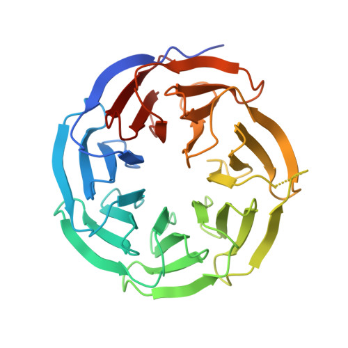
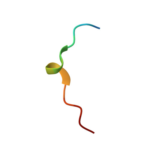
Structural Analysis of the Kansl1/Wdr5/Kansl2 Complex Reveals that Wdr5 is Required for Efficient Assembly and Chromatin Targeting of the Nsl Complex.

Dias, J.Van Nguyen, N.Georgiev, P.Gaub, A.Brettschneider, J.Cusack, S.Kadlec, J.Akhtar, A.

(2014) Genes Dev 28: 929

  • DOI: https://doi.org/10.1101/gad.240200.114
  • Primary Citation of Related Structures:  
    4CY1, 4CY2, 4CY3, 4CY5

  • PubMed Abstract: 

    The subunits of the nonspecific lethal (NSL) complex, which include the histone acetyltransferase MOF (males absent on the first), play important roles in various cellular functions, including transcription regulation and stem cell identity maintenance and reprogramming, and are frequently misregulated in disease. Here, we provide the first biochemical and structural insights into the molecular architecture of this large multiprotein assembly. We identified several direct interactions within the complex and show that KANSL1 acts as a scaffold protein interacting with four other subunits, including WDR5, which in turn binds KANSL2. Structural analysis of the KANSL1/WDR5/KANSL2 subcomplex reveals how WDR5 is recruited into the NSL complex via conserved linear motifs of KANSL1 and KANSL2. Using structure-based KANSL1 mutants in transgenic flies, we show that the KANSL1-WDR5 interaction is required for proper assembly, efficient recruitment of the NSL complex to target promoters, and fly viability. Our data clearly show that the interactions of WDR5 with the MOF-containing NSL complex and MLL/COMPASS histone methyltransferase complexes are mutually exclusive. We propose that rather than being a shared subunit, WDR5 plays an important role in assembling distinct histone-modifying complexes with different epigenetic regulatory roles.


  • Organizational Affiliation

    Grenoble Outstation, European Molecular Biology Laboratory (EMBL), 38042 Grenoble, France;


Macromolecules
Find similar proteins by:  (by identity cutoff)  |  3D Structure
Entity ID: 1
MoleculeChains Sequence LengthOrganismDetailsImage
WD REPEAT-CONTAINING PROTEIN 5
A, B
316Homo sapiensMutation(s): 0 
UniProt & NIH Common Fund Data Resources
Find proteins for P61964 (Homo sapiens)
Explore P61964 
Go to UniProtKB:  P61964
PHAROS:  P61964
GTEx:  ENSG00000196363 
Entity Groups  
Sequence Clusters30% Identity50% Identity70% Identity90% Identity95% Identity100% Identity
UniProt GroupP61964
Sequence Annotations
Expand
  • Reference Sequence

Find similar proteins by:  Sequence   |   3D Structure  

Entity ID: 2
MoleculeChains Sequence LengthOrganismDetailsImage
KAT8 REGULATORY NSL COMPLEX SUBUNIT 1
C, D
15Homo sapiensMutation(s): 0 
UniProt & NIH Common Fund Data Resources
Find proteins for Q7Z3B3 (Homo sapiens)
Explore Q7Z3B3 
Go to UniProtKB:  Q7Z3B3
PHAROS:  Q7Z3B3
GTEx:  ENSG00000120071 
Entity Groups  
Sequence Clusters30% Identity50% Identity70% Identity90% Identity95% Identity100% Identity
UniProt GroupQ7Z3B3
Sequence Annotations
Expand
  • Reference Sequence
Experimental Data & Validation

Experimental Data

  • Method: X-RAY DIFFRACTION
  • Resolution: 1.50 Å
  • R-Value Free: 0.183 
  • R-Value Work: 0.170 
  • R-Value Observed: 0.171 
  • Space Group: P 1 21 1
Unit Cell:
Length ( Å )Angle ( ˚ )
a = 39.76α = 90
b = 92.45β = 90.18
c = 81.17γ = 90
Software Package:
Software NamePurpose
REFMACrefinement
XDSdata reduction
XSCALEdata scaling
PHASERphasing

Structure Validation

View Full Validation Report


Entry History 

Deposition Data

Revision History  (Full details and data files)

  • Version 1.0: 2014-05-14
    Type: Initial release
  • Version 1.1: 2023-12-20
    Changes: Data collection, Database references, Derived calculations, Other, Refinement description