4CUZ

Crystal structure of S. aureus FabI in complex with NADPH and PT173


Experimental Data Snapshot

  • Method: X-RAY DIFFRACTION
  • Resolution: 3.10 Å
  • R-Value Free: 0.258 
  • R-Value Work: 0.207 
  • R-Value Observed: 0.209 

wwPDB Validation   3D Report Full Report


Ligand Structure Quality Assessment 


This is version 1.2 of the entry. See complete history


Literature

Rational Design of Broad Spectrum Antibacterial Activity Based on a Clinically Relevant Enoyl-Acyl Carrier Protein (Acp) Reductase Inhibitor.

Schiebel, J.Chang, A.Shah, S.Lu, Y.Liu, L.Pan, P.Hirschbeck, M.W.Tareilus, M.Eltschkner, S.Yu, W.Cummings, J.E.Knudson, S.E.Bommineni, G.R.Walker, S.G.Slayden, R.A.Sotriffer, C.A.Tonge, P.J.Kisker, C.

(2014) J Biol Chem 289: 15987

  • DOI: https://doi.org/10.1074/jbc.M113.532804
  • Primary Citation of Related Structures:  
    4BKU, 4CUZ, 4CV0, 4CV1, 4CV2, 4CV3

  • PubMed Abstract: 

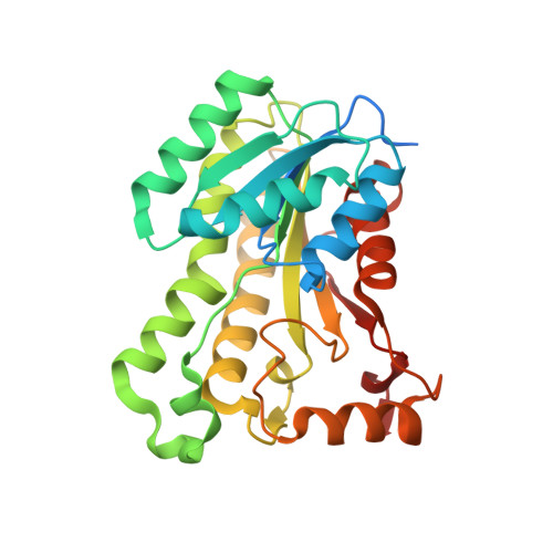
    Determining the molecular basis for target selectivity is of particular importance in drug discovery. The ideal antibiotic should be active against a broad spectrum of pathogenic organisms with a minimal effect on human targets. CG400549, a Staphylococcus-specific 2-pyridone compound that inhibits the enoyl-acyl carrier protein reductase (FabI), has recently been shown to possess human efficacy for the treatment of methicillin-resistant Staphylococcus aureus infections, which constitute a serious threat to human health. In this study, we solved the structures of three different FabI homologues in complex with several pyridone inhibitors, including CG400549. Based on these structures, we rationalize the 65-fold reduced affinity of CG400549 toward Escherichia coli versus S. aureus FabI and implement concepts to improve the spectrum of antibacterial activity. The identification of different conformational states along the reaction coordinate of the enzymatic hydride transfer provides an elegant visual depiction of the relationship between catalysis and inhibition, which facilitates rational inhibitor design. Ultimately, we developed the novel 4-pyridone-based FabI inhibitor PT166 that retained favorable pharmacokinetics and efficacy in a mouse model of S. aureus infection with extended activity against Gram-negative and mycobacterial organisms.


  • Organizational Affiliation

    From the Rudolf Virchow Center for Experimental Biomedicine, Institute for Structural Biology, University of Wuerzburg, D-97080 Wuerzburg, Germany, the Institute of Pharmacy and Food Chemistry, University of Wuerzburg, Am Hubland, D-97074 Wuerzburg, Germany.


Macromolecules
Find similar proteins by:  (by identity cutoff)  |  3D Structure
Entity ID: 1
MoleculeChains Sequence LengthOrganismDetailsImage
ENOYL-ACP REDUCTASE MOLECULE ENOYL-[ACYL-CARRIER-PROTEIN] REDUCTASE [NADPH]
A, B, C, D, E
A, B, C, D, E, F, G, H
282Staphylococcus aureusMutation(s): 1 
EC: 1.3.1.10
UniProt
Find proteins for A0A0H3JLH9 (Staphylococcus aureus (strain N315))
Explore A0A0H3JLH9 
Go to UniProtKB:  A0A0H3JLH9
Entity Groups  
Sequence Clusters30% Identity50% Identity70% Identity90% Identity95% Identity100% Identity
UniProt GroupA0A0H3JLH9
Sequence Annotations
Expand
  • Reference Sequence
Small Molecules
Ligands 3 Unique
IDChains Name / Formula / InChI Key2D Diagram3D Interactions
NDP
Query on NDP

Download Ideal Coordinates CCD File 
J [auth A]
L [auth B]
O [auth C]
Q [auth D]
S [auth E]
J [auth A],
L [auth B],
O [auth C],
Q [auth D],
S [auth E],
U [auth F],
W [auth G],
X [auth H]
NADPH DIHYDRO-NICOTINAMIDE-ADENINE-DINUCLEOTIDE PHOSPHATE
C21 H30 N7 O17 P3
ACFIXJIJDZMPPO-NNYOXOHSSA-N
AEW
Query on AEW

Download Ideal Coordinates CCD File 
I [auth A]
K [auth B]
N [auth C]
P [auth D]
R [auth E]
I [auth A],
K [auth B],
N [auth C],
P [auth D],
R [auth E],
T [auth F],
V [auth G]
1-(3-amino-2-methylbenzyl)-4-hexylpyridin-2(1H)-one
C19 H26 N2 O
PJADLCYVDLAZCT-UHFFFAOYSA-N
SO4
Query on SO4

Download Ideal Coordinates CCD File 
M [auth C]SULFATE ION
O4 S
QAOWNCQODCNURD-UHFFFAOYSA-L
Binding Affinity Annotations 
IDSourceBinding Affinity
AEW BindingDB:  4CUZ Ki: 2 (nM) from 1 assay(s)
Experimental Data & Validation

Experimental Data

  • Method: X-RAY DIFFRACTION
  • Resolution: 3.10 Å
  • R-Value Free: 0.258 
  • R-Value Work: 0.207 
  • R-Value Observed: 0.209 
  • Space Group: P 21 21 21
Unit Cell:
Length ( Å )Angle ( ˚ )
a = 61.48α = 90
b = 109.18β = 90
c = 289.371γ = 90
Software Package:
Software NamePurpose
PHENIXrefinement
iMOSFLMdata reduction
SCALAdata scaling
PHASERphasing

Structure Validation

View Full Validation Report


Ligand Structure Quality Assessment 


Entry History 

Deposition Data

Revision History  (Full details and data files)

  • Version 1.0: 2014-04-16
    Type: Initial release
  • Version 1.1: 2014-04-30
    Changes: Database references
  • Version 1.2: 2014-08-13
    Changes: Database references