The Cd27L and Ctp1L Endolysins Targeting Clostridia Contain a Built-in Trigger and Release Factor.
Dunne, M., Mertens, H.D.T., Garefalaki, V., Jeffries, C.M., Thompson, A., Lemke, E.A., Svergun, D.I., Mayer, M.J., Narbad, A., Meijers, R.(2014) PLoS Pathog 10: 04228
- PubMed: 25058163
- DOI: https://doi.org/10.1371/journal.ppat.1004228
- Primary Citation of Related Structures:
4CU2, 4CU5 - PubMed Abstract:
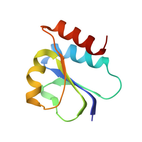
The bacteriophage ΦCD27 is capable of lysing Clostridium difficile, a pathogenic bacterium that is a major cause for nosocomial infection. A recombinant CD27L endolysin lyses C. difficile in vitro, and represents a promising alternative as a bactericide. To better understand the lysis mechanism, we have determined the crystal structure of an autoproteolytic fragment of the CD27L endolysin. The structure covers the C-terminal domain of the endolysin, and represents a novel fold that is identified in a number of lysins that target Clostridia bacteria. The structure indicates endolysin cleavage occurs at the stem of the linker connecting the catalytic domain with the C-terminal domain. We also solved the crystal structure of the C-terminal domain of a slow cleaving mutant of the CTP1L endolysin that targets C. tyrobutyricum. Two distinct dimerization modes are observed in the crystal structures for both endolysins, despite a sequence identity of only 22% between the domains. The dimers are validated to be present for the full length protein in solution by right angle light scattering, small angle X-ray scattering and cross-linking experiments using the cross-linking amino acid p-benzoyl-L-phenylalanine (pBpa). Mutagenesis on residues contributing to the dimer interfaces indicates that there is a link between the dimerization modes and the autocleavage mechanism. We show that for the CTP1L endolysin, there is a reduction in lysis efficiency that is proportional to the cleavage efficiency. We propose a model for endolysin triggering, where the extended dimer presents the inactive state, and a switch to the side-by-side dimer triggers the cleavage of the C-terminal domain. This leads to the release of the catalytic portion of the endolysin, enabling the efficient digestion of the bacterial cell wall.
Organizational Affiliation:
European Molecular Biology Laboratory (EMBL), Hamburg, Germany.