4CSV

Tyrosine kinase AS - a common ancestor of Src and Abl bound to Gleevec


Experimental Data Snapshot

  • Method: X-RAY DIFFRACTION
  • Resolution: 2.05 Å
  • R-Value Free: 0.242 
  • R-Value Work: 0.187 
  • R-Value Observed: 0.190 

wwPDB Validation   3D Report Full Report


Ligand Structure Quality Assessment 


This is version 1.2 of the entry. See complete history


Literature

Kinase Dynamics. Using Ancient Protein Kinases to Unravel a Modern Cancer Drug'S Mechanism.

Wilson, C.Agafonov, R.V.Hoemberger, M.Kutter, S.Zorba, A.Halpin, J.Buosi, V.Otten, R.Waterman, D.Theobald, D.L.Kern, D.

(2015) Science 347: 882

  • DOI: https://doi.org/10.1126/science.aaa1823
  • Primary Citation of Related Structures:  
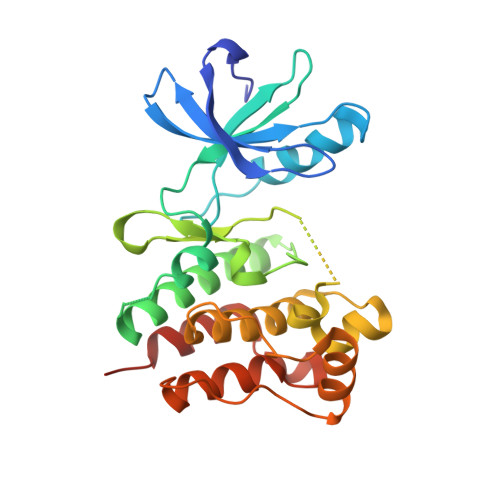
    4CSV, 4UEU

  • PubMed Abstract: 

    Macromolecular function is rooted in energy landscapes, where sequence determines not a single structure but an ensemble of conformations. Hence, evolution modifies a protein's function by altering its energy landscape. Here, we recreate the evolutionary pathway between two modern human oncogenes, Src and Abl, by reconstructing their common ancestors. Our evolutionary reconstruction combined with x-ray structures of the common ancestor and pre-steady-state kinetics reveals a detailed atomistic mechanism for selectivity of the successful cancer drug Gleevec. Gleevec affinity is gained during the evolutionary trajectory toward Abl and lost toward Src, primarily by shifting an induced-fit equilibrium that is also disrupted in the clinical T315I resistance mutation. This work reveals the mechanism of Gleevec specificity while offering insights into how energy landscapes evolve.


  • Organizational Affiliation

    Howard Hughes Medical Institute and Department of Biochemistry, Brandeis University, Waltham, MA 02452, USA.


Macromolecules
Find similar proteins by:  (by identity cutoff)  |  3D Structure
Entity ID: 1
MoleculeChains Sequence LengthOrganismDetailsImage
SRC-ABL TYROSINE KINASE ANCESTOR275synthetic constructMutation(s): 0 
EC: 2.7.10.2
Entity Groups  
Sequence Clusters30% Identity50% Identity70% Identity90% Identity95% Identity100% Identity
Sequence Annotations
Expand
  • Reference Sequence
Small Molecules
Ligands 1 Unique
IDChains Name / Formula / InChI Key2D Diagram3D Interactions
STI
Query on STI

Download Ideal Coordinates CCD File 
B [auth A]4-(4-METHYL-PIPERAZIN-1-YLMETHYL)-N-[4-METHYL-3-(4-PYRIDIN-3-YL-PYRIMIDIN-2-YLAMINO)-PHENYL]-BENZAMIDE
C29 H31 N7 O
KTUFNOKKBVMGRW-UHFFFAOYSA-N
Binding Affinity Annotations 
IDSourceBinding Affinity
STI Binding MOAD:  4CSV Ki: 910 (nM) from 1 assay(s)
Experimental Data & Validation

Experimental Data

  • Method: X-RAY DIFFRACTION
  • Resolution: 2.05 Å
  • R-Value Free: 0.242 
  • R-Value Work: 0.187 
  • R-Value Observed: 0.190 
  • Space Group: I 1 2 1
Unit Cell:
Length ( Å )Angle ( ˚ )
a = 71.029α = 90
b = 56.869β = 116.62
c = 76.118γ = 90
Software Package:
Software NamePurpose
REFMACrefinement
XDSdata reduction
Aimlessdata scaling
MOLREPphasing

Structure Validation

View Full Validation Report


Ligand Structure Quality Assessment 


Entry History 

Deposition Data

Revision History  (Full details and data files)

  • Version 1.0: 2015-03-04
    Type: Initial release
  • Version 1.1: 2019-05-08
    Changes: Data collection, Experimental preparation, Other
  • Version 1.2: 2023-12-20
    Changes: Data collection, Database references, Derived calculations, Other, Refinement description, Structure summary