4CNO

Structure of the Salmonella typhi Type I dehydroquinase inhibited by a 3-dehydroquinic acid derivative


Experimental Data Snapshot

  • Method: X-RAY DIFFRACTION
  • Resolution: 1.50 Å
  • R-Value Free: 0.214 
  • R-Value Work: 0.180 
  • R-Value Observed: 0.182 

wwPDB Validation   3D Report Full Report


Ligand Structure Quality Assessment 


This is version 1.2 of the entry. See complete history


Literature

Insights into substrate binding and catalysis in bacterial type I dehydroquinase.

Maneiro, M.Peon, A.Lence, E.Otero, J.M.Van Raaij, M.J.Thompson, P.Hawkins, A.R.Gonzalez-Bello, C.

(2014) Biochem J 462: 415-424

  • DOI: https://doi.org/10.1042/BJ20140614
  • Primary Citation of Related Structures:  
    4CNO

  • PubMed Abstract: 

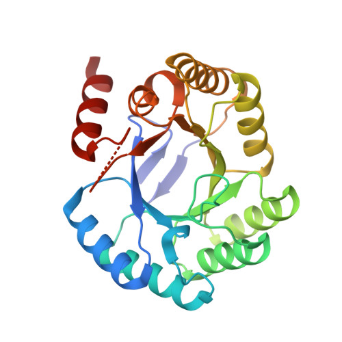
    Structural, biochemical and computational studies to study substrate binding and the role of the conserved residues of the DHQ1 (type I dehydroquinase) enzyme active site are reported in the present paper. The crystal structure of DHQ1 from Salmonella typhi in complex with (2R)-2-methyl-3-dehydroquinic acid, a substrate analogue, was solved at 1.5 Å. The present study reveals a previously unknown key role for conserved Glu46, Phe145 and Met205 and Gln236, Pro234 and Ala233 residues, with the latter three being located in the flexible substrate-covering loop. Gln236 was shown to be responsible for the folding of this loop and for the dramatic reduction of its flexibility, which triggers active site closure. Glu46 was found to be key in bringing the substrate close to the lysine/histidine catalytic pocket to initiate catalysis. The present study could be useful in the rational design of inhibitors of this challenging and recognized target for the development of novel herbicides and antimicrobial agents.


  • Organizational Affiliation

    *Centro Singular de Investigación en Química Biológica y Materiales Moleculares (CIQUS), Universidad de Santiago de Compostela, calle Jenaro de la Fuente s/n, 15782 Santiago de Compostela, Spain.


Macromolecules
Find similar proteins by:  (by identity cutoff)  |  3D Structure
Entity ID: 1
MoleculeChains Sequence LengthOrganismDetailsImage
3-DEHYDROQUINATE DEHYDRATASE
A, B, C, D
252Salmonella enterica subsp. enterica serovar TyphiMutation(s): 0 
EC: 4.2.1.10
UniProt
Find proteins for P24670 (Salmonella typhi)
Explore P24670 
Go to UniProtKB:  P24670
Entity Groups  
Sequence Clusters30% Identity50% Identity70% Identity90% Identity95% Identity100% Identity
UniProt GroupP24670
Sequence Annotations
Expand
  • Reference Sequence
Experimental Data & Validation

Experimental Data

  • Method: X-RAY DIFFRACTION
  • Resolution: 1.50 Å
  • R-Value Free: 0.214 
  • R-Value Work: 0.180 
  • R-Value Observed: 0.182 
  • Space Group: P 1 21 1
Unit Cell:
Length ( Å )Angle ( ˚ )
a = 78.67α = 90
b = 39.406β = 104.68
c = 155.13γ = 90
Software Package:
Software NamePurpose
REFMACrefinement
XDSdata reduction
SCALEdata scaling
MOLREPphasing

Structure Validation

View Full Validation Report


Ligand Structure Quality Assessment 


Entry History 

Deposition Data

Revision History  (Full details and data files)

  • Version 1.0: 2014-10-08
    Type: Initial release
  • Version 1.1: 2018-02-07
    Changes: Database references
  • Version 1.2: 2023-12-20
    Changes: Data collection, Database references, Derived calculations, Other, Refinement description