4CKL

Structure of 55 kDa N-terminal domain of E. coli DNA gyrase A subunit with simocyclinone D8 bound


Experimental Data Snapshot

  • Method: X-RAY DIFFRACTION
  • Resolution: 2.05 Å
  • R-Value Free: 0.225 
  • R-Value Work: 0.196 
  • R-Value Observed: 0.197 

wwPDB Validation   3D Report Full Report


Ligand Structure Quality Assessment 


This is version 1.4 of the entry. See complete history


Literature

A New Crystal Structure of the Bifunctional Antibiotic Simocyclinone D8 Bound to DNA Gyrase Gives Fresh Insight Into the Mechanism of Inhibition.

Hearnshaw, S.J.Edwards, M.J.Stevenson, C.E.Lawson, D.M.Maxwell, A.

(2014) J Mol Biol 426: 2023

  • DOI: https://doi.org/10.1016/j.jmb.2014.02.017
  • Primary Citation of Related Structures:  
    4CKK, 4CKL

  • PubMed Abstract: 

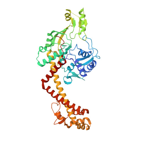
    Simocyclinone D8 (SD8) is an antibiotic produced by Streptomyces antibioticus that targets DNA gyrase. A previous structure of SD8 complexed with the N-terminal domain of the DNA gyrase A protein (GyrA) suggested that four SD8 molecules stabilized a tetramer of the protein; subsequent mass spectrometry experiments suggested that a protein dimer with two symmetry-related SD8s was more likely. This work describes the structures of a further truncated form of the GyrA N-terminal domain fragment with and without SD8 bound. The structure with SD8 has the two SD8 molecules bound within the same GyrA dimer. This new structure is entirely consistent with the mutations in GyrA that confer SD8 resistance and, by comparison with a new apo structure of the GyrA N-terminal domain, reveals the likely conformation changes that occur upon SD8 binding and the detailed mechanism of SD8 inhibition of gyrase. Isothermal titration calorimetry experiments are consistent with the crystallography results and further suggest that a previously observed complex between SD8 and GyrB is ~1000-fold weaker than the interaction with GyrA.


  • Organizational Affiliation

    Department of Biological Chemistry, John Innes Centre, Norwich Research Park, Norwich NR4 7UH, UK.


Macromolecules
Find similar proteins by:  (by identity cutoff)  |  3D Structure
Entity ID: 1
MoleculeChains Sequence LengthOrganismDetailsImage
DNA GYRASE SUBUNIT A
A, B
493Escherichia coliMutation(s): 0 
EC: 5.99.1.3
UniProt
Find proteins for A0A0H3JH39 (Escherichia coli O157:H7)
Explore A0A0H3JH39 
Go to UniProtKB:  A0A0H3JH39
Entity Groups  
Sequence Clusters30% Identity50% Identity70% Identity90% Identity95% Identity100% Identity
UniProt GroupA0A0H3JH39
Sequence Annotations
Expand
  • Reference Sequence
Small Molecules
Ligands 1 Unique
IDChains Name / Formula / InChI Key2D Diagram3D Interactions
SM8
Query on SM8

Download Ideal Coordinates CCD File 
C [auth A]SIMOCYCLINONE D8
C46 H42 Cl N O18
PLEGMCYXNQPJNV-VHSVXIAXSA-N
Binding Affinity Annotations 
IDSourceBinding Affinity
SM8 Binding MOAD:  4CKL Kd: 44 (nM) from 1 assay(s)
Experimental Data & Validation

Experimental Data

  • Method: X-RAY DIFFRACTION
  • Resolution: 2.05 Å
  • R-Value Free: 0.225 
  • R-Value Work: 0.196 
  • R-Value Observed: 0.197 
  • Space Group: C 1 2 1
Unit Cell:
Length ( Å )Angle ( ˚ )
a = 160.82α = 90
b = 96.05β = 132.72
c = 112.35γ = 90
Software Package:
Software NamePurpose
REFMACrefinement
XDSdata reduction
Aimlessdata scaling
PHASERphasing

Structure Validation

View Full Validation Report


Ligand Structure Quality Assessment 


Entry History 

Deposition Data

Revision History  (Full details and data files)

  • Version 1.0: 2014-03-12
    Type: Initial release
  • Version 1.1: 2014-03-19
    Changes: Database references
  • Version 1.2: 2014-04-16
    Changes: Database references
  • Version 1.3: 2014-05-14
    Changes: Database references
  • Version 1.4: 2023-12-20
    Changes: Data collection, Database references, Derived calculations, Other, Refinement description