4CJ7

Structure of Crenactin, an archeal actin-like protein


Experimental Data Snapshot

  • Method: X-RAY DIFFRACTION
  • Resolution: 3.20 Å
  • R-Value Free: 0.295 
  • R-Value Work: 0.235 
  • R-Value Observed: 0.238 

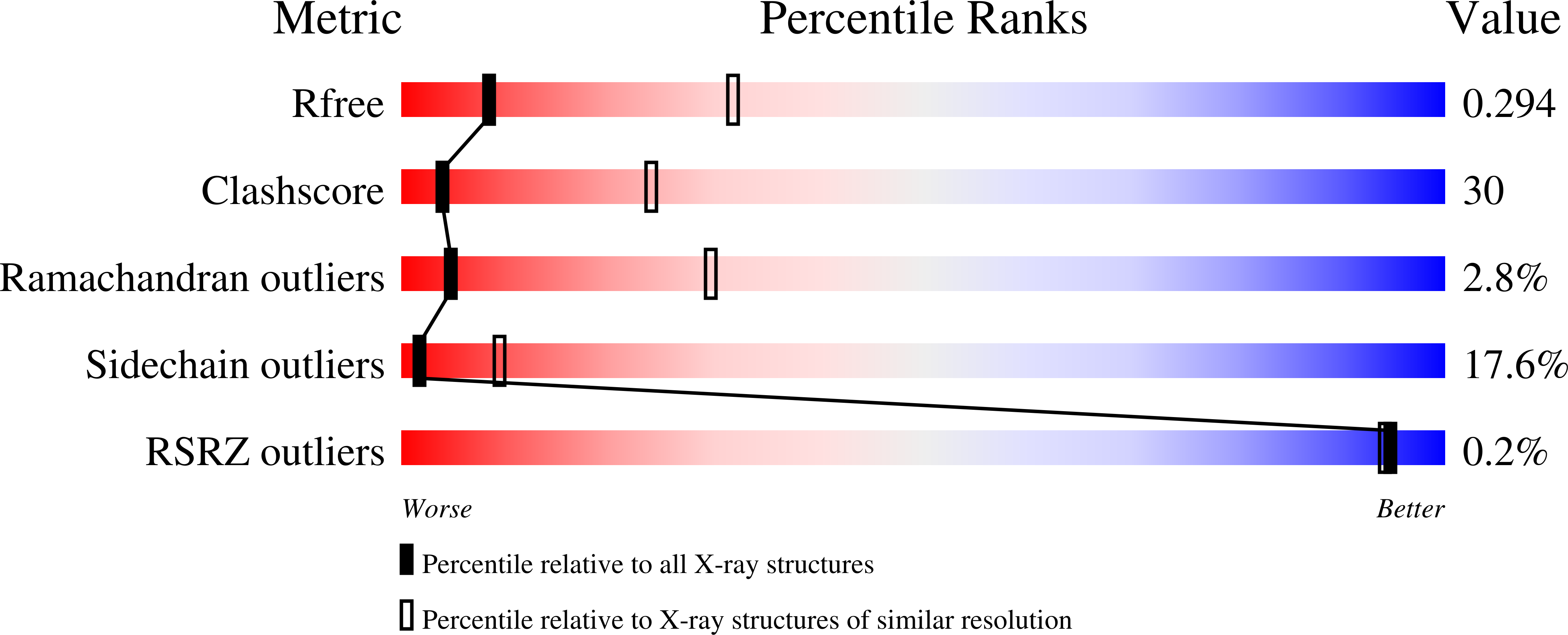
wwPDB Validation   3D Report Full Report


Ligand Structure Quality Assessment 


This is version 1.3 of the entry. See complete history


Literature

Crenactin from Pyrobaculum Calidifontis is Closely Related to Actin in Structure and Forms Steep Helical Filaments.

Izore, T.Duman, R.Kureisaite-Ciziene, D.Lowe, J.

(2014) FEBS Lett 588: 776

  • DOI: https://doi.org/10.1016/j.febslet.2014.01.029
  • Primary Citation of Related Structures:  
    4CJ7

  • PubMed Abstract: 

    Polymerising proteins of the actin family are nearly ubiquitous. Crenactins, restricted to Crenarchaea, are more closely related to actin than bacterial MreB. Crenactins occur in gene clusters hinting at an unknown, but conserved function. We solved the crystal structure of crenactin at 3.2 Å resolution. The protein crystallises as a continuous right-handed helix with 8 subunits per complete turn, spanning 419 Å. The structure of crenactin shows several loops that are longer than in actin, but overall, crenactin is closely related to eukaryotic actin, with an RMSD of 1.6 Å. Crenactin filaments imaged by electron microscopy showed polymers with very similar helical parameters.


  • Organizational Affiliation

    MRC Laboratory of Molecular Biology, Structural Studies Division, Francis Crick Avenue, Cambridge CB2 0QH, United Kingdom.


Macromolecules
Find similar proteins by:  (by identity cutoff)  |  3D Structure
Entity ID: 1
MoleculeChains Sequence LengthOrganismDetailsImage
ACTIN/ACTIN FAMILY PROTEIN
A, B
432Pyrobaculum calidifontis JCM 11548Mutation(s): 0 
UniProt
Find proteins for A3MWN5 (Pyrobaculum calidifontis (strain DSM 21063 / JCM 11548 / VA1))
Explore A3MWN5 
Go to UniProtKB:  A3MWN5
Entity Groups  
Sequence Clusters30% Identity50% Identity70% Identity90% Identity95% Identity100% Identity
UniProt GroupA3MWN5
Sequence Annotations
Expand
  • Reference Sequence
Small Molecules
Experimental Data & Validation

Experimental Data

  • Method: X-RAY DIFFRACTION
  • Resolution: 3.20 Å
  • R-Value Free: 0.295 
  • R-Value Work: 0.235 
  • R-Value Observed: 0.238 
  • Space Group: P 41 21 2
Unit Cell:
Length ( Å )Angle ( ˚ )
a = 78.127α = 90
b = 78.127β = 90
c = 418.958γ = 90
Software Package:
Software NamePurpose
REFMACrefinement
XDSdata reduction
SCALAdata scaling
PHASERphasing

Structure Validation

View Full Validation Report


Ligand Structure Quality Assessment 


Entry History 

Deposition Data

Revision History  (Full details and data files)

  • Version 1.0: 2014-01-22
    Type: Initial release
  • Version 1.1: 2014-02-12
    Changes: Database references
  • Version 1.2: 2014-03-05
    Changes: Database references
  • Version 1.3: 2023-12-20
    Changes: Data collection, Database references, Derived calculations, Other, Refinement description