4CIU

Crystal structure of E. coli ClpB


Experimental Data Snapshot

  • Method: X-RAY DIFFRACTION
  • Resolution: 3.50 Å
  • R-Value Free: 0.247 
  • R-Value Work: 0.224 
  • R-Value Observed: 0.225 

wwPDB Validation   3D Report Full Report


Ligand Structure Quality Assessment 


This is version 1.2 of the entry. See complete history


Literature

Head-to-Tail Interactions of the Coiled-Coil Domains Regulate Clpb Cooperation with Hsp70 in Protein Disaggregation

Carroni, M.Kummer, E.Oguchi, Y.Wendler, P.Clare, D.K.Sinning, I.Kopp, J.Mogk, A.Bukau, B.Saibil, H.

(2014) Elife 3: 02481

  • DOI: https://doi.org/10.7554/eLife.02481
  • Primary Citation of Related Structures:  
    4CIU, 4D2Q, 4D2U, 4D2X

  • PubMed Abstract: 

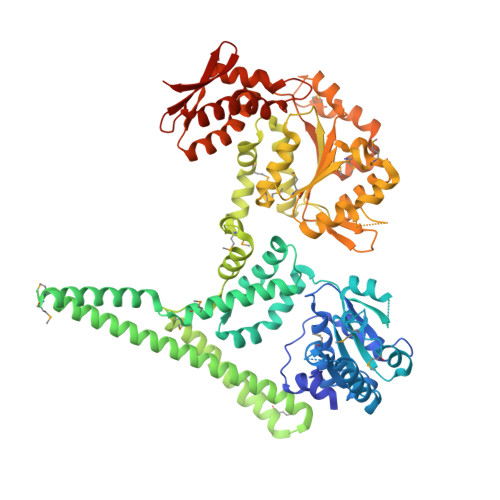
    The hexameric AAA+ chaperone ClpB reactivates aggregated proteins in cooperation with the Hsp70 system. Essential for disaggregation, the ClpB middle domain (MD) is a coiled-coil propeller that binds Hsp70. Although the ClpB subunit structure is known, positioning of the MD in the hexamer and its mechanism of action are unclear. We obtained electron microscopy (EM) structures of the BAP variant of ClpB that binds the protease ClpP, clearly revealing MD density on the surface of the ClpB ring. Mutant analysis and asymmetric reconstructions show that MDs adopt diverse positions in a single ClpB hexamer. Adjacent, horizontally oriented MDs form head-to-tail contacts and repress ClpB activity by preventing Hsp70 interaction. Tilting of the MD breaks this contact, allowing Hsp70 binding, and releasing the contact in adjacent subunits. Our data suggest a wavelike activation of ClpB subunits around the ring.DOI: http://dx.doi.org/10.7554/eLife.02481.001.


  • Organizational Affiliation

    Department of Crystallography, Birkbeck College, University of London, London, United Kingdom.


Macromolecules
Find similar proteins by:  (by identity cutoff)  |  3D Structure
Entity ID: 1
MoleculeChains Sequence LengthOrganismDetailsImage
CHAPERONE PROTEIN CLPB727Escherichia coliMutation(s): 3 
UniProt
Find proteins for P63284 (Escherichia coli (strain K12))
Explore P63284 
Go to UniProtKB:  P63284
Entity Groups  
Sequence Clusters30% Identity50% Identity70% Identity90% Identity95% Identity100% Identity
UniProt GroupP63284
Sequence Annotations
Expand
  • Reference Sequence
Small Molecules
Modified Residues  1 Unique
IDChains TypeFormula2D DiagramParent
MSE
Query on MSE
A
L-PEPTIDE LINKINGC5 H11 N O2 SeMET
Experimental Data & Validation

Experimental Data

  • Method: X-RAY DIFFRACTION
  • Resolution: 3.50 Å
  • R-Value Free: 0.247 
  • R-Value Work: 0.224 
  • R-Value Observed: 0.225 
  • Space Group: P 65
Unit Cell:
Length ( Å )Angle ( ˚ )
a = 127.329α = 90
b = 127.329β = 90
c = 119.852γ = 120
Software Package:
Software NamePurpose
PHENIXrefinement
MOSFLMdata reduction
SCALAdata scaling
PHENIXphasing

Structure Validation

View Full Validation Report


Ligand Structure Quality Assessment 


Entry History 

Deposition Data

Revision History  (Full details and data files)

  • Version 1.0: 2014-05-14
    Type: Initial release
  • Version 1.1: 2014-05-28
    Changes: Database references
  • Version 1.2: 2019-05-22
    Changes: Data collection, Derived calculations, Experimental preparation, Other