4CH2

Low-salt crystal structure of a thrombin-GpIbalpha peptide complex


Experimental Data Snapshot

  • Method: X-RAY DIFFRACTION
  • Resolution: 1.60 Å
  • R-Value Free: 0.187 
  • R-Value Work: 0.155 
  • R-Value Observed: 0.156 

wwPDB Validation   3D Report Full Report


Ligand Structure Quality Assessment 


This is version 1.4 of the entry. See complete history


Literature

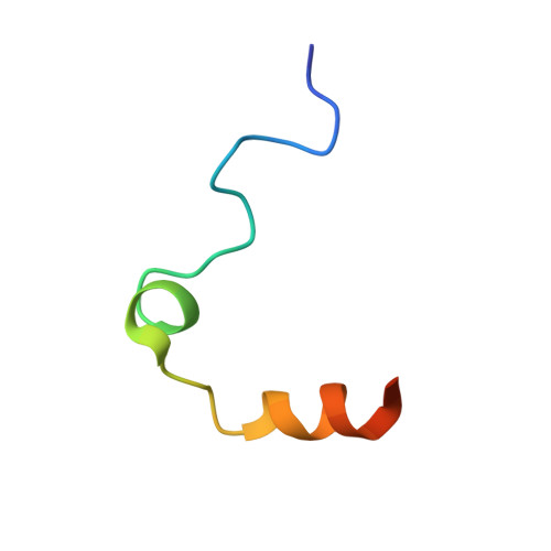
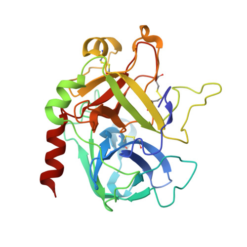
Gpibalpha Interacts Exclusively with Exosite II of Thrombin

Lechtenberg, B.C.Freund, S.M.V.Huntington, J.A.

(2014) J Mol Biol 426: 881

  • DOI: https://doi.org/10.1016/j.jmb.2013.11.027
  • Primary Citation of Related Structures:  
    4CH2, 4CH8

  • PubMed Abstract: 

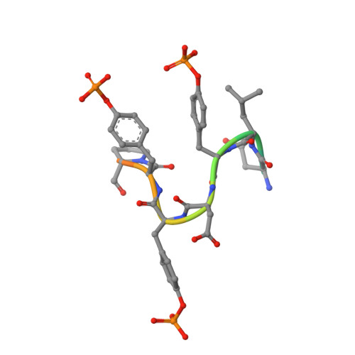
    Activation of platelets by the serine protease thrombin is a critical event in haemostasis. This process involves the binding of thrombin to glycoprotein Ibα (GpIbα) and cleavage of protease-activated receptors (PARs). The N-terminal extracellular domain of GpIbα contains an acidic peptide stretch that has been identified as the main thrombin binding site, and both anion binding exosites of thrombin have been implicated in GpIbα binding, but it remains unclear how they are involved. This issue is of critical importance for the mechanism of platelet activation by thrombin. If both exosites bind to GpIbα, thrombin could potentially act as a platelet adhesion molecule or receptor dimerisation trigger. Alternatively, if only a single site is involved, GpIbα may serve as a cofactor for PAR-1 activation by thrombin. To determine the involvement of thrombin's two exosites in GpIbα binding, we employed the complementary methods of mutational analysis, binding studies, X-ray crystallography and NMR spectroscopy. Our results indicate that the peptide corresponding to the C-terminal portion of GpIbα and the entire extracellular domain bind exclusively to thrombin's exosite II. The interaction of thrombin with GpIbα thus serves to recruit thrombin activity to the platelet surface while leaving exosite I free for PAR-1 recognition.


  • Organizational Affiliation

    Department of Haematology, Cambridge Institute for Medical Research, University of Cambridge, Wellcome Trust/MRC Building, Hills Road, Cambridge CB2 0XY, United Kingdom.


Macromolecules
Find similar proteins by:  (by identity cutoff)  |  3D Structure
Entity ID: 1
MoleculeChains Sequence LengthOrganismDetailsImage
THROMBIN, LIGHT CHAIN
A, C
36Homo sapiensMutation(s): 0 
EC: 3.4.21.5
UniProt & NIH Common Fund Data Resources
Find proteins for P00734 (Homo sapiens)
Explore P00734 
Go to UniProtKB:  P00734
PHAROS:  P00734
GTEx:  ENSG00000180210 
Entity Groups  
Sequence Clusters30% Identity50% Identity70% Identity90% Identity95% Identity100% Identity
UniProt GroupP00734
Sequence Annotations
Expand
  • Reference Sequence
Find similar proteins by:  (by identity cutoff)  |  3D Structure
Entity ID: 2
MoleculeChains Sequence LengthOrganismDetailsImage
THROMBIN, HEAVY CHAIN
B, D
259Homo sapiensMutation(s): 0 
EC: 3.4.21.5
UniProt & NIH Common Fund Data Resources
Find proteins for P00734 (Homo sapiens)
Explore P00734 
Go to UniProtKB:  P00734
PHAROS:  P00734
GTEx:  ENSG00000180210 
Entity Groups  
Sequence Clusters30% Identity50% Identity70% Identity90% Identity95% Identity100% Identity
UniProt GroupP00734
Sequence Annotations
Expand
  • Reference Sequence

Find similar proteins by:  Sequence   |   3D Structure  

Entity ID: 3
MoleculeChains Sequence LengthOrganismDetailsImage
PLATELET GLYCOPROTEIN IB ALPHA CHAIN, RESIDUES 287-300E [auth P],
F [auth Q]
14Homo sapiensMutation(s): 0 
UniProt & NIH Common Fund Data Resources
Find proteins for P07359 (Homo sapiens)
Explore P07359 
Go to UniProtKB:  P07359
PHAROS:  P07359
GTEx:  ENSG00000185245 
Entity Groups  
Sequence Clusters30% Identity50% Identity70% Identity90% Identity95% Identity100% Identity
UniProt GroupP07359
Sequence Annotations
Expand
  • Reference Sequence
Small Molecules
Ligands 3 Unique
IDChains Name / Formula / InChI Key2D Diagram3D Interactions
0G6
Query on 0G6

Download Ideal Coordinates CCD File 
H [auth B],
K [auth D]
D-phenylalanyl-N-[(2S,3S)-6-{[amino(iminio)methyl]amino}-1-chloro-2-hydroxyhexan-3-yl]-L-prolinamide
C21 H34 Cl N6 O3
DVFLYEYCMMLBTQ-VSZNYVQBSA-O
GOL
Query on GOL

Download Ideal Coordinates CCD File 
G [auth A],
I [auth B],
L [auth D],
M [auth D]
GLYCEROL
C3 H8 O3
PEDCQBHIVMGVHV-UHFFFAOYSA-N
NA
Query on NA

Download Ideal Coordinates CCD File 
J [auth B],
N [auth D]
SODIUM ION
Na
FKNQFGJONOIPTF-UHFFFAOYSA-N
Modified Residues  1 Unique
IDChains TypeFormula2D DiagramParent
PTR
Query on PTR
E [auth P],
F [auth Q]
L-PEPTIDE LINKINGC9 H12 N O6 PTYR
Biologically Interesting Molecules (External Reference) 1 Unique
Experimental Data & Validation

Experimental Data

  • Method: X-RAY DIFFRACTION
  • Resolution: 1.60 Å
  • R-Value Free: 0.187 
  • R-Value Work: 0.155 
  • R-Value Observed: 0.156 
  • Space Group: C 1 2 1
Unit Cell:
Length ( Å )Angle ( ˚ )
a = 149.57α = 90
b = 50.56β = 97.15
c = 76.28γ = 90
Software Package:
Software NamePurpose
REFMACrefinement
MOSFLMdata reduction
SCALAdata scaling
PHASERphasing

Structure Validation

View Full Validation Report


Ligand Structure Quality Assessment 


Entry History 

Deposition Data

Revision History  (Full details and data files)

  • Version 1.0: 2013-12-11
    Type: Initial release
  • Version 1.1: 2013-12-18
    Changes: Database references
  • Version 1.2: 2013-12-25
    Changes: Non-polymer description
  • Version 1.3: 2014-02-12
    Changes: Database references
  • Version 1.4: 2023-12-20
    Changes: Data collection, Database references, Derived calculations, Other, Refinement description