4CEJ

Crystal structure of AddAB-DNA-ADPNP complex at 3 Angstrom resolution


Experimental Data Snapshot

  • Method: X-RAY DIFFRACTION
  • Resolution: 3.00 Å
  • R-Value Free: 0.243 
  • R-Value Work: 0.203 
  • R-Value Observed: 0.205 

wwPDB Validation   3D Report Full Report


Ligand Structure Quality Assessment 


This is version 1.3 of the entry. See complete history


Literature

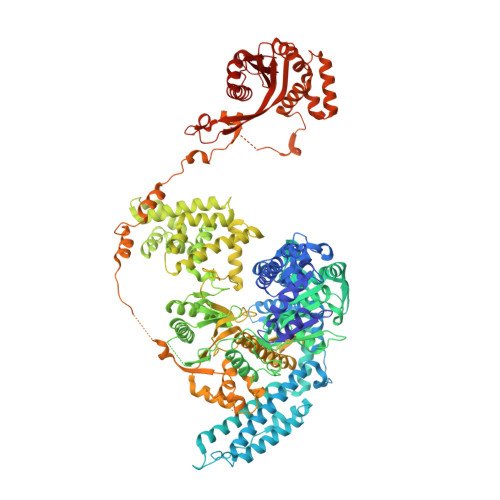
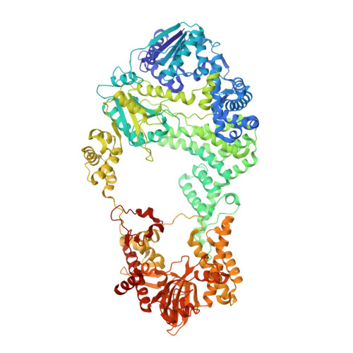
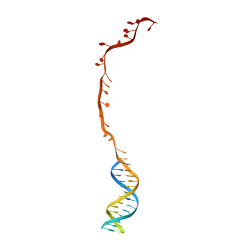
Structural Basis for Translocation by Addab Helicase-Nuclease and its Arrest at Chi Sites.

Krajewski, W.W.Fu, X.Wilkinson, M.Cronin, N.B.Dillingham, M.S.Wigley, D.B.

(2014) Nature 508: 416

  • DOI: https://doi.org/10.1038/nature13037

  • PubMed Abstract: 

    In bacterial cells, processing of double-stranded DNA breaks for repair by homologous recombination is dependent upon the recombination hotspot sequence χ (Chi) and is catalysed by either an AddAB- or RecBCD-type helicase-nuclease (reviewed in refs 3, 4). These enzyme complexes unwind and digest the DNA duplex from the broken end until they encounter a χ sequence, whereupon they produce a 3' single-stranded DNA tail onto which they initiate loading of the RecA protein. Consequently, regulation of the AddAB/RecBCD complex by χ is a key control point in DNA repair and other processes involving genetic recombination. Here we report crystal structures of Bacillus subtilis AddAB in complex with different χ-containing DNA substrates either with or without a non-hydrolysable ATP analogue. Comparison of these structures suggests a mechanism for DNA translocation and unwinding, suggests how the enzyme binds specifically to χ sequences, and explains how χ recognition leads to the arrest of AddAB (and RecBCD) translocation that is observed in single-molecule experiments.


  • Organizational Affiliation

    1] Division of Structural Biology, Institute of Cancer Research, Chester Beatty Laboratories, 237 Fulham Road, London SW3 6JB, UK [2] CRT Discovery Laboratories, Department of Biological Sciences, Birkbeck, University of London, London WC1E 7HX, UK.


Macromolecules

Find similar proteins by:  (by identity cutoff)  |  3D Structure
Entity ID: 1
MoleculeChains Sequence LengthOrganismDetailsImage
ATP-DEPENDENT HELICASE/NUCLEASE SUBUNIT A1,232Bacillus subtilis subsp. subtilis str. 168Mutation(s): 2 
EC: 3.1 (PDB Primary Data), 3.6.4.12 (PDB Primary Data)
UniProt
Find proteins for P23478 (Bacillus subtilis (strain 168))
Explore P23478 
Go to UniProtKB:  P23478
Entity Groups  
Sequence Clusters30% Identity50% Identity70% Identity90% Identity95% Identity100% Identity
UniProt GroupP23478
Sequence Annotations
Expand
  • Reference Sequence
Find similar proteins by:  (by identity cutoff)  |  3D Structure
Entity ID: 2
MoleculeChains Sequence LengthOrganismDetailsImage
ATP-DEPENDENT HELICASE/DEOXYRIBONUCLEASE SUBUNIT B1,166Bacillus subtilis subsp. subtilis str. 168Mutation(s): 3 
EC: 3.1 (PDB Primary Data), 3.6.4.12 (PDB Primary Data)
UniProt
Find proteins for P23477 (Bacillus subtilis (strain 168))
Explore P23477 
Go to UniProtKB:  P23477
Entity Groups  
Sequence Clusters30% Identity50% Identity70% Identity90% Identity95% Identity100% Identity
UniProt GroupP23477
Sequence Annotations
Expand
  • Reference Sequence
Find similar nucleic acids by:  (by identity cutoff)  |  3D Structure
Entity ID: 3
MoleculeChains LengthOrganismImage
DNAC [auth X]70synthetic construct
Sequence Annotations
Expand
  • Reference Sequence
Experimental Data & Validation

Experimental Data

  • Method: X-RAY DIFFRACTION
  • Resolution: 3.00 Å
  • R-Value Free: 0.243 
  • R-Value Work: 0.203 
  • R-Value Observed: 0.205 
  • Space Group: P 1 21 1
Unit Cell:
Length ( Å )Angle ( ˚ )
a = 77.437α = 90
b = 152.941β = 94.3
c = 125.244γ = 90
Software Package:
Software NamePurpose
PHENIXrefinement
XDSdata reduction
SCALAdata scaling
PHENIXphasing

Structure Validation

View Full Validation Report


Ligand Structure Quality Assessment 


Entry History 

Deposition Data

Revision History  (Full details and data files)

  • Version 1.0: 2014-03-12
    Type: Initial release
  • Version 1.1: 2014-04-09
    Changes: Database references
  • Version 1.2: 2014-04-23
    Changes: Database references
  • Version 1.3: 2023-12-20
    Changes: Data collection, Database references, Derived calculations, Other, Refinement description