


4CA8
Drosophila Angiotensin converting enzyme (AnCE) in complex with a phosphinic tripeptide FII
- PDB DOI: https://doi.org/10.2210/pdb4CA8/pdb
- Classification: HYDROLASE
- Organism(s): Drosophila melanogaster
- Expression System: Komagataella pastoris
- Mutation(s): No
- Deposited: 2013-10-07 Released: 2013-12-11
Experimental Data Snapshot
- Method: X-RAY DIFFRACTION
- Resolution: 1.99 Å
- R-Value Free: 0.216
- R-Value Work: 0.194
- R-Value Observed: 0.195

This is version 2.1 of the entry. See complete history.

Macromolecules
Find similar proteins by:
(by identity cutoff) | 3D Structure
Entity ID: 1 | |||||
|---|---|---|---|---|---|
| Molecule | Chains | Sequence Length | Organism | Details | Image |
| ANGIOTENSIN-CONVERTING ENZYME | 598 | Drosophila melanogaster | Mutation(s): 0 EC: 3.4.15.1 | 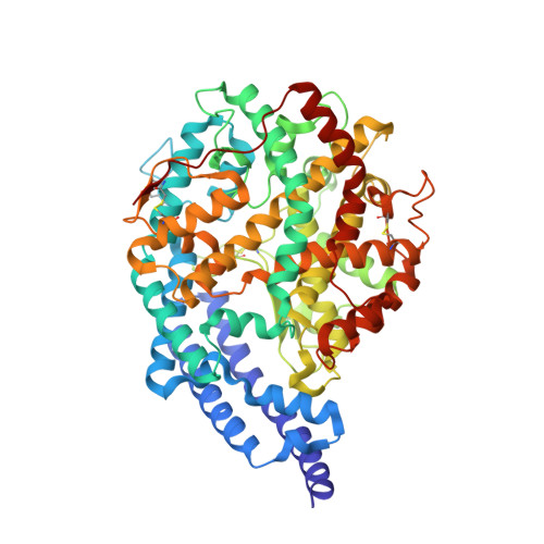 | |
UniProt | |||||
Find proteins for Q10714 (Drosophila melanogaster) Explore Q10714 Go to UniProtKB: Q10714 | |||||
Entity Groups | |||||
| Sequence Clusters | 30% Identity50% Identity70% Identity90% Identity95% Identity100% Identity | ||||
| UniProt Group | Q10714 | ||||
Sequence AnnotationsExpand | |||||
| |||||
Oligosaccharides
Entity ID: 2 | |||||
|---|---|---|---|---|---|
| Molecule | Chains | Length | 2D Diagram | Glycosylation | 3D Interactions |
| beta-D-mannopyranose-(1-6)-alpha-D-mannopyranose-(1-3)-[alpha-D-mannopyranose-(1-6)]beta-D-mannopyranose-(1-4)-2-acetamido-2-deoxy-beta-D-glucopyranose-(1-4)-2-acetamido-2-deoxy-beta-D-glucopyranose | B | 6 |  | N-Glycosylation | |
Glycosylation Resources | |||||
GlyTouCan: G64803HZ GlyCosmos: G64803HZ GlyGen: G64803HZ | |||||
Small Molecules
| Ligands 4 Unique | |||||
|---|---|---|---|---|---|
| ID | Chains | Name / Formula / InChI Key | 2D Diagram | 3D Interactions | |
| 3ES Query on 3ES | C [auth A] | [(2S)-2-({3-[HYDROXYL(2-PHENYL-(1R)-1-{[(BENZYLOXY)[(2S)-2-({3-[HYDROXYL(2-PHENYL-(1R)-1-CARBONYL]-AMINO}ETHYL)PHOSPHINYL]-2-[(3-PHENYLISOXAZOL-5-YL)METHYL]-1-OXO-PROPYL}AMINO)-3-(4-HYDROXY-PHENYL) C38 H38 N3 O9 P ZPFSKFCSVXPMBD-PFESQZPFSA-N |  | ||
| NAG Query on NAG | I [auth A], J [auth A] | 2-acetamido-2-deoxy-beta-D-glucopyranose C8 H15 N O6 OVRNDRQMDRJTHS-FMDGEEDCSA-N |  | ||
| SO4 Query on SO4 | D [auth A], E [auth A], F [auth A], G [auth A] | SULFATE ION O4 S QAOWNCQODCNURD-UHFFFAOYSA-L |  | ||
| ZN Query on ZN | H [auth A] | ZINC ION Zn PTFCDOFLOPIGGS-UHFFFAOYSA-N |  | ||
Experimental Data & Validation
Experimental Data
- Method: X-RAY DIFFRACTION
- Resolution: 1.99 Å
- R-Value Free: 0.216
- R-Value Work: 0.194
- R-Value Observed: 0.195
- Space Group: H 3
Unit Cell:
| Length ( Å ) | Angle ( ˚ ) |
|---|---|
| a = 172.89 | α = 90 |
| b = 172.89 | β = 90 |
| c = 100.48 | γ = 120 |
| Software Name | Purpose |
|---|---|
| REFMAC | refinement |
| MOSFLM | data reduction |
| SCALA | data scaling |
| PHASER | phasing |
Entry History
Deposition Data
- Released Date: 2013-12-11 Deposition Author(s): Masuyer, G., Akif, M., Czarny, B., Beau, F., Schwager, S.L.U., Sturrock, E.D., Isaac, R.E., Dive, V., Acharya, K.R.
Revision History (Full details and data files)
- Version 1.0: 2013-12-11
Type: Initial release - Version 1.1: 2014-02-12
Changes: Database references - Version 2.0: 2020-07-29
Type: Remediation
Reason: Carbohydrate remediation
Changes: Atomic model, Data collection, Derived calculations, Other, Structure summary - Version 2.1: 2023-12-20
Changes: Data collection, Database references, Refinement description, Structure summary
