4C5L

Structure of the pyridoxal kinase from Staphylococcus aureus in complex with pyridoxal


Experimental Data Snapshot

  • Method: X-RAY DIFFRACTION
  • Resolution: 1.85 Å
  • R-Value Free: 0.275 
  • R-Value Work: 0.234 
  • R-Value Observed: 0.236 

wwPDB Validation   3D Report Full Report


Ligand Structure Quality Assessment 


This is version 1.3 of the entry. See complete history


Literature

A Subfamily of Bacterial Ribokinases Utilizes a Hemithioacetal for Pyridoxal Phosphate Salvage.

Nodwell, M.B.Koch, M.F.Alte, F.Schneider, S.Sieber, S.A.

(2014) J Am Chem Soc 136: 4992

  • DOI: https://doi.org/10.1021/ja411785r
  • Primary Citation of Related Structures:  
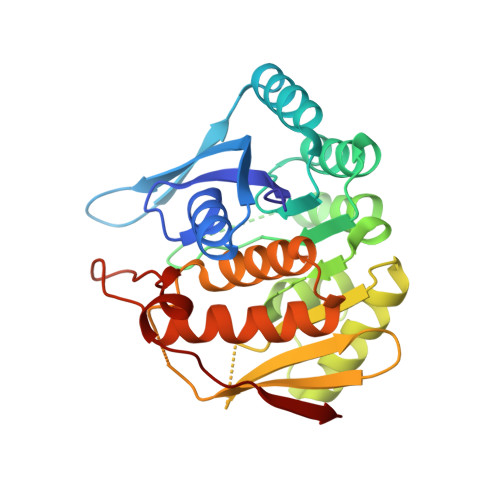
    4C5J, 4C5K, 4C5L, 4C5M, 4C5N

  • PubMed Abstract: 

    Pyridoxal 5'-phosphate (PLP) is the active vitamer of vitamin B6 and acts as an essential cofactor in many aspects of amino acid and sugar metabolism. The virulence and survival of pathogenic bacteria such as Mycobacterium tuberculosis depend on PLP, and deficiencies in humans have also been associated with neurological disorders and inflammation. While PLP can be synthesized by a de novo pathway in bacteria and plants, most higher organisms rely on a salvage pathway that phosphorylates either pyridoxal (PL) or its related vitamers, pyridoxine (PN) and pyridoxamine (PM). PL kinases (PLKs) are essential for this phosphorylation step and are thus of major importance for cellular viability. We recently identified a pyridoxal kinase (SaPLK) as a target of the natural product antibiotic rugulactone (Ru) in Staphylococcus aureus. Surprisingly, Ru selectively modified SaPLK not at the active site cysteine, but on a remote cysteine residue. Based on structural and biochemical studies, we now provide insight into an unprecedented dual Cys charge relay network that is mandatory for PL phosphorylation. The key component is the reactive Cys 110 residue in the lid region that forms a hemithioactetal intermediate with the 4'-aldehyde of PL. This hemithioacetal, in concert with the catalytic Cys 214, increases the nucleophilicity of the PL 5'-OH group for the inline displacement reaction with the γ-phosphate of ATP. A closer inspection of related enzymes reveals that Cys 110 is conserved and thus serves as a characteristic mechanistic feature for a dual-function ribokinase subfamily herein termed CC-PLKs.


  • Organizational Affiliation

    Organic Chemistry II, Centre for Integrated Protein Science CIPSM, Institute of Advanced Studies, and ‡Biochemistry, Department of Chemistry, Technische Universität München , Lichtenbergstrasse 4, 85747 Garching, Germany.


Macromolecules
Find similar proteins by:  (by identity cutoff)  |  3D Structure
Entity ID: 1
MoleculeChains Sequence LengthOrganismDetailsImage
PHOSPHOMETHYLPYRIMIDINE KINASE
A, B, C, D
276Staphylococcus aureus subsp. aureus Mu50Mutation(s): 0 
EC: 2.7.1.35
UniProt
Find proteins for A0A0H3JTP0 (Staphylococcus aureus (strain Mu50 / ATCC 700699))
Explore A0A0H3JTP0 
Go to UniProtKB:  A0A0H3JTP0
Entity Groups  
Sequence Clusters30% Identity50% Identity70% Identity90% Identity95% Identity100% Identity
UniProt GroupA0A0H3JTP0
Sequence Annotations
Expand
  • Reference Sequence
Small Molecules
Ligands 3 Unique
IDChains Name / Formula / InChI Key2D Diagram3D Interactions
UEG
Query on UEG

Download Ideal Coordinates CCD File 
E [auth A],
J [auth C],
K [auth D]
4,5-bis(hydroxymethyl)-2-methyl-pyridin-3-ol
C8 H11 N O3
LXNHXLLTXMVWPM-UHFFFAOYSA-N
PXL
Query on PXL

Download Ideal Coordinates CCD File 
H [auth B]3-HYDROXY-5-(HYDROXYMETHYL)-2-METHYLISONICOTINALDEHYDE
C8 H9 N O3
RADKZDMFGJYCBB-UHFFFAOYSA-N
SO4
Query on SO4

Download Ideal Coordinates CCD File 
F [auth A],
G [auth A],
I [auth B],
L [auth D]
SULFATE ION
O4 S
QAOWNCQODCNURD-UHFFFAOYSA-L
Experimental Data & Validation

Experimental Data

  • Method: X-RAY DIFFRACTION
  • Resolution: 1.85 Å
  • R-Value Free: 0.275 
  • R-Value Work: 0.234 
  • R-Value Observed: 0.236 
  • Space Group: P 1 21 1
Unit Cell:
Length ( Å )Angle ( ˚ )
a = 63.44α = 90
b = 102.78β = 90.44
c = 83.52γ = 90
Software Package:
Software NamePurpose
REFMACrefinement
XDSdata reduction
XSCALEdata scaling

Structure Validation

View Full Validation Report


Ligand Structure Quality Assessment 


Entry History 

Deposition Data

Revision History  (Full details and data files)

  • Version 1.0: 2014-03-19
    Type: Initial release
  • Version 1.1: 2014-04-16
    Changes: Database references
  • Version 1.2: 2019-10-16
    Changes: Data collection, Derived calculations, Other
  • Version 1.3: 2023-12-20
    Changes: Data collection, Database references, Derived calculations, Refinement description