4C4K

Crystal structure of the titin M10-Obscurin Ig domain 1 complex


Experimental Data Snapshot

  • Method: X-RAY DIFFRACTION
  • Resolution: 1.95 Å
  • R-Value Free: 0.210 
  • R-Value Work: 0.171 
  • R-Value Observed: 0.173 

wwPDB Validation   3D Report Full Report


This is version 1.3 of the entry. See complete history


Literature

The Crystal Structure of the Human Titin:Obscurin Complex Reveals a Conserved Yet Specific Muscle M-Band Zipper Module.

Pernigo, S.Fukuzawa, A.Pandini, A.Holt, M.Kleinjung, J.Gautel, M.Steiner, R.A.

(2015) J Mol Biol 427: 718

  • DOI: https://doi.org/10.1016/j.jmb.2014.11.019
  • Primary Citation of Related Structures:  
    4C4K, 4UOW

  • PubMed Abstract: 

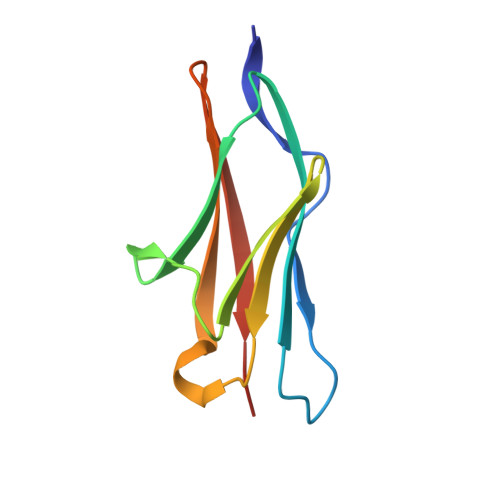
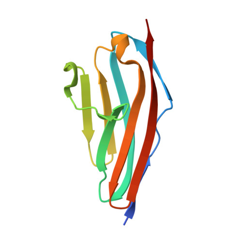
    M10 is the most C-terminal immunoglobulin (Ig) domain of the giant protein titin and a frequent target of disease-linked mutations. Currently, it is the only known muscle Ig domain able to interact with two alternative ligands-obscurin and obscurin-like-1 (Obsl1)-in different sarcomeric subregions. Obscurin and Obsl1 use their homologous N-terminal Ig domain (O1 in obscurin and OL1 in Obsl1) to bind M10 in a mutually exclusive manner. We present here the X-ray structure of the human titin:obscurin M10:O1 complex extending our previous work on the M10:OL1 interaction. Similar to M10:OL1, the M10:O1 complex displays a chevron-shaped antiparallel Ig-Ig architecture held together by a conserved molecular interface, which we validated by isothermal titration calorimetry and sorting experiments in neonatal rat cardiomyocytes. O1, although structurally related to OL1 and M10, both members of the intermediate set (I-set) Ig family, presents an intriguing switch of its βA' strand. This leads to structural differences between the complexes, particularly for the "open side" of the chevron-shaped assembly. A bioinformatics analysis reveals that the βA'-switch observed for O1 is rare and that it is involved in mediating protein-protein interactions. Molecular dynamics simulations also suggest that this topological alteration substantially increases local flexibility compared to the conventional I-set Ig domains. The O1/OL1 Ig domains are candidate discriminatory structural modules potentially directing the binding of specific additional partners at the M-band. Cellular sorting experiments in neonatal rat cardiomyocytes are consistent with the view that the titin:obscurin/Obsl1 complexes might be a platform for higher-order interactions.


  • Organizational Affiliation

    Randall Division of Cell and Molecular Biophysics, King's College London, New Hunt's House, Guy's Campus, London SE1 1UL, UK.


Macromolecules
Find similar proteins by:  (by identity cutoff)  |  3D Structure
Entity ID: 1
MoleculeChains Sequence LengthOrganismDetailsImage
OBSCURINA [auth O]98Homo sapiensMutation(s): 0 
EC: 2.7.11.1
UniProt & NIH Common Fund Data Resources
Find proteins for Q5VST9 (Homo sapiens)
Explore Q5VST9 
Go to UniProtKB:  Q5VST9
PHAROS:  Q5VST9
GTEx:  ENSG00000154358 
Entity Groups  
Sequence Clusters30% Identity50% Identity70% Identity90% Identity95% Identity100% Identity
UniProt GroupQ5VST9
Sequence Annotations
Expand
  • Reference Sequence
Find similar proteins by:  (by identity cutoff)  |  3D Structure
Entity ID: 2
MoleculeChains Sequence LengthOrganismDetailsImage
TITINB [auth T]102Homo sapiensMutation(s): 0 
EC: 2.7.11.1
UniProt & NIH Common Fund Data Resources
Find proteins for Q8WZ42 (Homo sapiens)
Explore Q8WZ42 
Go to UniProtKB:  Q8WZ42
PHAROS:  Q8WZ42
GTEx:  ENSG00000155657 
Entity Groups  
Sequence Clusters30% Identity50% Identity70% Identity90% Identity95% Identity100% Identity
UniProt GroupQ8WZ42
Sequence Annotations
Expand
  • Reference Sequence
Small Molecules
Ligands 1 Unique
IDChains Name / Formula / InChI Key2D Diagram3D Interactions
EDO
Query on EDO

Download Ideal Coordinates CCD File 
C [auth O]
D [auth O]
E [auth O]
F [auth O]
G [auth O]
C [auth O],
D [auth O],
E [auth O],
F [auth O],
G [auth O],
H [auth O],
I [auth O],
J [auth T],
K [auth T],
L [auth T],
M [auth T],
N [auth T]
1,2-ETHANEDIOL
C2 H6 O2
LYCAIKOWRPUZTN-UHFFFAOYSA-N
Experimental Data & Validation

Experimental Data

  • Method: X-RAY DIFFRACTION
  • Resolution: 1.95 Å
  • R-Value Free: 0.210 
  • R-Value Work: 0.171 
  • R-Value Observed: 0.173 
  • Space Group: P 21 21 21
Unit Cell:
Length ( Å )Angle ( ˚ )
a = 40.94α = 90
b = 66.88β = 90
c = 72.93γ = 90
Software Package:
Software NamePurpose
BUSTERrefinement
XDSdata reduction
SCALAdata scaling
MOLREPphasing

Structure Validation

View Full Validation Report


Entry History 

Deposition Data

Revision History  (Full details and data files)

  • Version 1.0: 2014-09-24
    Type: Initial release
  • Version 1.1: 2014-12-17
    Changes: Database references
  • Version 1.2: 2015-03-04
    Changes: Database references
  • Version 1.3: 2023-12-20
    Changes: Data collection, Database references, Derived calculations, Other, Refinement description