4C2V

Aurora B kinase in complex with the specific inhibitor Barasertib


Experimental Data Snapshot

  • Method: X-RAY DIFFRACTION
  • Resolution: 1.49 Å
  • R-Value Free: 0.197 
  • R-Value Work: 0.167 
  • R-Value Observed: 0.168 

wwPDB Validation   3D Report Full Report


Ligand Structure Quality Assessment 


This is version 1.2 of the entry. See complete history


Literature

Structure of Aurora B-Incenp in Complex with Barasertib Reveals a Potential Transinhibitory Mechanism

Sessa, F.Villa, F.

(2014) Acta Crystallogr Sect F Struct Biol Cryst Commun 70: 294

  • DOI: https://doi.org/10.1107/S2053230X14002118
  • Primary Citation of Related Structures:  
    4C2V, 4C2W

  • PubMed Abstract: 

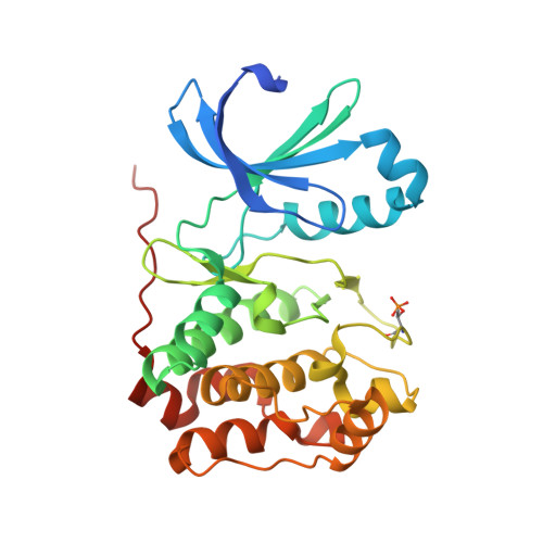
    The Aurora family is a well conserved and well characterized group of serine-threonine kinases involved in the normal progression of mitosis. The deregulation of Aurora kinases impairs spindle assembly, checkpoint function and cell division. To date, many small molecules that compete with ATP for binding to Aurora kinases have been developed and characterized. Here, the first structure of the Xenopus laevis Aurora B-INCENP complex bound to the clinically relevant small molecule barasertib was determined. The binding properties of this inhibitor to the Aurora B active site are analyzed and reported. An unexpected crystal-packing contact in the Aurora B-INCENP structure coordinated by an ATP analogue is also reported, in which the INCENP C-terminus occupies the substrate-binding region, resembling the protein kinase A inhibitory mechanism.


  • Organizational Affiliation

    Department of Experimental Oncology, European Institute of Oncology, Via Adamello 16, 20139 Milan, Italy.


Macromolecules
Find similar proteins by:  (by identity cutoff)  |  3D Structure
Entity ID: 1
MoleculeChains Sequence LengthOrganismDetailsImage
AURORA KINASE B-A
A, B
285Xenopus laevisMutation(s): 0 
EC: 2.7.11.1
UniProt
Find proteins for Q6DE08 (Xenopus laevis)
Explore Q6DE08 
Go to UniProtKB:  Q6DE08
Entity Groups  
Sequence Clusters30% Identity50% Identity70% Identity90% Identity95% Identity100% Identity
UniProt GroupQ6DE08
Sequence Annotations
Expand
  • Reference Sequence
Find similar proteins by:  (by identity cutoff)  |  3D Structure
Entity ID: 2
MoleculeChains Sequence LengthOrganismDetailsImage
INNER CENTROMERE PROTEIN A
C, D
44Xenopus laevisMutation(s): 0 
UniProt
Find proteins for O13024 (Xenopus laevis)
Explore O13024 
Go to UniProtKB:  O13024
Entity Groups  
Sequence Clusters30% Identity50% Identity70% Identity90% Identity95% Identity100% Identity
UniProt GroupO13024
Sequence Annotations
Expand
  • Reference Sequence
Small Molecules
Ligands 1 Unique
IDChains Name / Formula / InChI Key2D Diagram3D Interactions
YJA
Query on YJA

Download Ideal Coordinates CCD File 
E [auth A],
F [auth B]
2-[5-[[7-[3-[ethyl(2-hydroxyethyl)amino]propoxy]quinazolin-4-yl]amino]-1H-pyrazol-3-yl]-N-(3-fluorophenyl)ethanamide
C26 H30 F N7 O3
QYZOGCMHVIGURT-UHFFFAOYSA-N
Modified Residues  1 Unique
IDChains TypeFormula2D DiagramParent
TPO
Query on TPO
A, B
L-PEPTIDE LINKINGC4 H10 N O6 PTHR
Binding Affinity Annotations 
IDSourceBinding Affinity
YJA Binding MOAD:  4C2V Ki: 1 (nM) from 1 assay(s)
Experimental Data & Validation

Experimental Data

  • Method: X-RAY DIFFRACTION
  • Resolution: 1.49 Å
  • R-Value Free: 0.197 
  • R-Value Work: 0.167 
  • R-Value Observed: 0.168 
  • Space Group: P 1 21 1
Unit Cell:
Length ( Å )Angle ( ˚ )
a = 46.065α = 90
b = 68.101β = 96.65
c = 117.087γ = 90
Software Package:
Software NamePurpose
REFMACrefinement
XDSdata reduction
SCALAdata scaling
MOLREPphasing

Structure Validation

View Full Validation Report


Ligand Structure Quality Assessment 


Entry History 

Deposition Data

Revision History  (Full details and data files)

  • Version 1.0: 2014-02-26
    Type: Initial release
  • Version 1.1: 2014-03-12
    Changes: Database references
  • Version 1.2: 2014-03-19
    Changes: Database references