4C2L

Crystal structure of endo-xylogalacturonan hydrolase from Aspergillus tubingensis


Experimental Data Snapshot

  • Method: X-RAY DIFFRACTION
  • Resolution: 1.75 Å
  • R-Value Free: 0.189 
  • R-Value Work: 0.167 
  • R-Value Observed: 0.168 

wwPDB Validation   3D Report Full Report


Ligand Structure Quality Assessment 


This is version 2.1 of the entry. See complete history


Literature

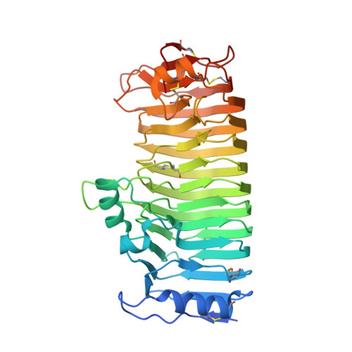
Crystal Structure of Endo-Xylogalacturonan Hydrolase from Aspergillus Tubingensis.

Rozeboom, H.J.Beldman, G.Schols, H.A.Dijkstra, B.W.

(2013) FEBS J 280: 6061

  • DOI: https://doi.org/10.1111/febs.12524
  • Primary Citation of Related Structures:  
    4C2L

  • PubMed Abstract: 

    Endo-xylogalacturonan hydrolase is a member of glycoside hydrolase family 28 (GH28) that hydrolyzes the glycosidic bond between two β-xylose-substituted galacturonic acid residues in pectin. Presented here is the X-ray crystal structure of the endo-xylogalacturonan hydrolase from Aspergillus tubingensis (XghA) at 1.75 Å resolution. The high degree of structural conservation in the active site and catalytic apparatus compared with polygalacturonases indicates that cleavage of the substrate proceeds in essentially the same way as found for the other GH28 enzymes. Molecular modeling of a xylosylated tri-galacturonate in the active site identified the amino acid residues involved in substrate binding. They border a substrate-binding cleft that is much wider than in other polygalacturonases, and can accommodate xylosylated substrates. The most extensive interactions appear to occur at subsite +2, in agreement with the enzyme kinetics results, which showed enhanced activity on substrates with a xylose attached to the galacturonic acid bound at subsite +2.


  • Organizational Affiliation

    Laboratory of Biophysical Chemistry, Groningen Biomolecular Sciences and Biotechnology Institute, University of Groningen, The Netherlands.


Macromolecules
Find similar proteins by:  (by identity cutoff)  |  3D Structure
Entity ID: 1
MoleculeChains Sequence LengthOrganismDetailsImage
ENDO-XYLOGALACTURONAN HYDROLASE A388Aspergillus tubingensisMutation(s): 0 
EC: 3.2.1
UniProt
Find proteins for Q9UUZ2 (Aspergillus tubingensis)
Explore Q9UUZ2 
Go to UniProtKB:  Q9UUZ2
Entity Groups  
Sequence Clusters30% Identity50% Identity70% Identity90% Identity95% Identity100% Identity
UniProt GroupQ9UUZ2
Sequence Annotations
Expand
  • Reference Sequence
Oligosaccharides

Help

Entity ID: 2
MoleculeChains Length2D Diagram Glycosylation3D Interactions
2-acetamido-2-deoxy-beta-D-glucopyranose-(1-4)-2-acetamido-2-deoxy-beta-D-glucopyranose
B
2N-Glycosylation
Glycosylation Resources
GlyTouCan:  G42666HT
GlyCosmos:  G42666HT
GlyGen:  G42666HT
Small Molecules
Ligands 4 Unique
IDChains Name / Formula / InChI Key2D Diagram3D Interactions
NAG
Query on NAG

Download Ideal Coordinates CCD File 
D [auth A]2-acetamido-2-deoxy-beta-D-glucopyranose
C8 H15 N O6
OVRNDRQMDRJTHS-FMDGEEDCSA-N
MAN
Query on MAN

Download Ideal Coordinates CCD File 
C [auth A]alpha-D-mannopyranose
C6 H12 O6
WQZGKKKJIJFFOK-PQMKYFCFSA-N
SO4
Query on SO4

Download Ideal Coordinates CCD File 
E [auth A]
F [auth A]
G [auth A]
H [auth A]
I [auth A]
E [auth A],
F [auth A],
G [auth A],
H [auth A],
I [auth A],
J [auth A],
K [auth A]
SULFATE ION
O4 S
QAOWNCQODCNURD-UHFFFAOYSA-L
GOL
Query on GOL

Download Ideal Coordinates CCD File 
L [auth A]GLYCEROL
C3 H8 O3
PEDCQBHIVMGVHV-UHFFFAOYSA-N
Experimental Data & Validation

Experimental Data

  • Method: X-RAY DIFFRACTION
  • Resolution: 1.75 Å
  • R-Value Free: 0.189 
  • R-Value Work: 0.167 
  • R-Value Observed: 0.168 
  • Space Group: I 2 2 2
Unit Cell:
Length ( Å )Angle ( ˚ )
a = 76.44α = 90
b = 121.18β = 90
c = 129.67γ = 90
Software Package:
Software NamePurpose
REFMACrefinement
iMOSFLMdata reduction
SCALAdata scaling
PHASERphasing

Structure Validation

View Full Validation Report


Ligand Structure Quality Assessment 


Entry History 

Deposition Data

Revision History  (Full details and data files)

  • Version 1.0: 2013-09-25
    Type: Initial release
  • Version 1.1: 2013-12-04
    Changes: Database references
  • Version 1.2: 2019-01-30
    Changes: Data collection, Experimental preparation, Other
  • Version 1.3: 2019-02-27
    Changes: Data collection, Derived calculations, Experimental preparation
  • Version 2.0: 2020-07-29
    Type: Remediation
    Reason: Carbohydrate remediation
    Changes: Atomic model, Data collection, Derived calculations, Other, Structure summary
  • Version 2.1: 2023-12-20
    Changes: Data collection, Database references, Refinement description, Structure summary