4C0X

The crystal strucuture of PpAzoR in complex with anthraquinone-2- sulfonate


Experimental Data Snapshot

  • Method: X-RAY DIFFRACTION
  • Resolution: 1.50 Å
  • R-Value Free: 0.193 
  • R-Value Work: 0.169 
  • R-Value Observed: 0.170 

wwPDB Validation   3D Report Full Report


Ligand Structure Quality Assessment 


This is version 1.3 of the entry. See complete history


Literature

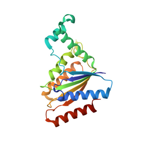
The Crystal Structure of Pseudomonas Putida Azor: The Active Site Revisited.

Goncalves, A.M.D.Mendes, S.De Sanctis, D.Martins, L.O.Bento, I.

(2013) FEBS J 280: 6643

  • DOI: https://doi.org/10.1111/febs.12568
  • Primary Citation of Related Structures:  
    4C0W, 4C0X, 4C14

  • PubMed Abstract: 

    The enzymatic degradation of azo dyes begins with the reduction of the azo bond. In this article, we report the crystal structures of the native azoreductase from Pseudomonas putida MET94 (PpAzoR) (1.60 Å), of PpAzoR in complex with anthraquinone-2-sulfonate (1.50 Å), and of PpAzoR in complex with Reactive Black 5 dye (1.90 Å). These structures reveal the residues and subtle changes that accompany substrate binding and release. Such changes highlight the fine control of access to the catalytic site that is required by the ping-pong mechanism, and in turn the specificity offered by the enzyme towards different substrates. The topology surrounding the active site shows novel features of substrate recognition and binding that help to explain and differentiate the substrate specificity observed among different bacterial azoreductases.


  • Organizational Affiliation

    Macromolecular Crystallography Unit, Instituto de Tecnologia Química e Biológica, Universidade Nova de Lisboa, Oeiras, Portugal.


Macromolecules
Find similar proteins by:  (by identity cutoff)  |  3D Structure
Entity ID: 1
MoleculeChains Sequence LengthOrganismDetailsImage
FMN-DEPENDENT NADH-AZOREDUCTASE 1203Pseudomonas putidaMutation(s): 0 
EC: 1.7 (PDB Primary Data), 1.6.5.2 (PDB Primary Data)
UniProt
Find proteins for Q88IY3 (Pseudomonas putida (strain ATCC 47054 / DSM 6125 / CFBP 8728 / NCIMB 11950 / KT2440))
Explore Q88IY3 
Go to UniProtKB:  Q88IY3
Entity Groups  
Sequence Clusters30% Identity50% Identity70% Identity90% Identity95% Identity100% Identity
UniProt GroupQ88IY3
Sequence Annotations
Expand
  • Reference Sequence
Small Molecules
Ligands 4 Unique
IDChains Name / Formula / InChI Key2D Diagram3D Interactions
FMN
Query on FMN

Download Ideal Coordinates CCD File 
B [auth A]FLAVIN MONONUCLEOTIDE
C17 H21 N4 O9 P
FVTCRASFADXXNN-SCRDCRAPSA-N
AQN
Query on AQN

Download Ideal Coordinates CCD File 
C [auth A]9,10-dioxo-9,10-dihydroanthracene-2-sulfonic acid
C14 H8 O5 S
MMNWSHJJPDXKCH-UHFFFAOYSA-N
PG4
Query on PG4

Download Ideal Coordinates CCD File 
F [auth A],
G [auth A]
TETRAETHYLENE GLYCOL
C8 H18 O5
UWHCKJMYHZGTIT-UHFFFAOYSA-N
GOL
Query on GOL

Download Ideal Coordinates CCD File 
D [auth A],
E [auth A]
GLYCEROL
C3 H8 O3
PEDCQBHIVMGVHV-UHFFFAOYSA-N
Experimental Data & Validation

Experimental Data

  • Method: X-RAY DIFFRACTION
  • Resolution: 1.50 Å
  • R-Value Free: 0.193 
  • R-Value Work: 0.169 
  • R-Value Observed: 0.170 
  • Space Group: F 2 2 2
Unit Cell:
Length ( Å )Angle ( ˚ )
a = 72.712α = 90
b = 94.947β = 90
c = 146.511γ = 90
Software Package:
Software NamePurpose
PHENIXrefinement
iMOSFLMdata reduction
SCALAdata scaling
MOLREPphasing

Structure Validation

View Full Validation Report


Ligand Structure Quality Assessment 


Entry History 

Deposition Data

Revision History  (Full details and data files)

  • Version 1.0: 2013-10-30
    Type: Initial release
  • Version 1.1: 2013-12-18
    Changes: Database references
  • Version 1.2: 2017-08-09
    Changes: Data collection
  • Version 1.3: 2023-12-20
    Changes: Data collection, Database references, Derived calculations, Other, Refinement description