4BWP

Structure of Drosophila Melanogaster PAN3 pseudokinase


Experimental Data Snapshot

  • Method: X-RAY DIFFRACTION
  • Resolution: 3.60 Å
  • R-Value Free: 0.269 
  • R-Value Work: 0.220 
  • R-Value Observed: 0.222 

wwPDB Validation   3D Report Full Report


Ligand Structure Quality Assessment 


This is version 1.2 of the entry. See complete history


Literature

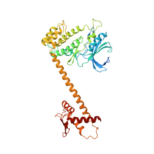
Structure of the Pan3 Pseudokinase Reveals the Basis for Interactions with the Pan2 Deadenylase and the Gw182 Proteins

Christie, M.Boland, A.Huntzinger, E.Weichenrieder, O.Izaurralde, E.

(2013) Mol Cell 51: 360

  • DOI: https://doi.org/10.1016/j.molcel.2013.07.011
  • Primary Citation of Related Structures:  
    4BWK, 4BWP, 4BWX

  • PubMed Abstract: 

    The PAN2-PAN3 deadenylase complex functions in general and miRNA-mediated mRNA degradation and is specifically recruited to miRNA targets by GW182/TNRC6 proteins. We describe the PAN3 adaptor protein crystal structure that, unexpectedly, forms intertwined and asymmetric homodimers. Dimerization is mediated by a coiled coil that links an N-terminal pseudokinase to a C-terminal knob domain. The PAN3 pseudokinase binds ATP, and this function is required for mRNA degradation in vivo. We further identified conserved surfaces required for mRNA degradation, including the binding surface for the PAN2 deadenylase on the knob domain. The most remarkable structural feature is the presence of a tryptophan-binding pocket at the dimer interface, which mediates binding to TNRC6C in human cells. Together, our data reveal the structural basis for the interaction of PAN3 with PAN2 and the recruitment of the PAN2-PAN3 complex to miRNA targets by TNRC6 proteins.


  • Organizational Affiliation

    Department of Biochemistry, Max Planck Institute for Developmental Biology, Spemannstrasse 35, 72076 Tübingen, Germany.


Macromolecules
Find similar proteins by:  (by identity cutoff)  |  3D Structure
Entity ID: 1
MoleculeChains Sequence LengthOrganismDetailsImage
PAB-DEPENDENT POLY(A)-SPECIFIC RIBONUCLEASE SUBUNIT PAN-3
A, B
448Drosophila melanogasterMutation(s): 0 
UniProt
Find proteins for Q95RR8 (Drosophila melanogaster)
Explore Q95RR8 
Go to UniProtKB:  Q95RR8
Entity Groups  
Sequence Clusters30% Identity50% Identity70% Identity90% Identity95% Identity100% Identity
UniProt GroupQ95RR8
Sequence Annotations
Expand
  • Reference Sequence
Small Molecules
Experimental Data & Validation

Experimental Data

  • Method: X-RAY DIFFRACTION
  • Resolution: 3.60 Å
  • R-Value Free: 0.269 
  • R-Value Work: 0.220 
  • R-Value Observed: 0.222 
  • Space Group: P 43 2 2
Unit Cell:
Length ( Å )Angle ( ˚ )
a = 102.171α = 90
b = 102.171β = 90
c = 264.715γ = 90
Software Package:
Software NamePurpose
REFMACrefinement
XDSdata reduction
SCALAdata scaling
PHASERphasing

Structure Validation

View Full Validation Report


Ligand Structure Quality Assessment 


Entry History 

Deposition Data

Revision History  (Full details and data files)

  • Version 1.0: 2013-08-21
    Type: Initial release
  • Version 1.1: 2013-08-28
    Changes: Database references
  • Version 1.2: 2023-12-20
    Changes: Data collection, Database references, Derived calculations, Other, Refinement description