4BW9

PylRS Y306G, Y384F, I405R mutant in complex with AMP-PNP


Experimental Data Snapshot

  • Method: X-RAY DIFFRACTION
  • Resolution: 2.35 Å
  • R-Value Free: 0.204 
  • R-Value Work: 0.174 
  • R-Value Observed: 0.176 

wwPDB Validation   3D Report Full Report


Ligand Structure Quality Assessment 


This is version 1.3 of the entry. See complete history


Literature

Structural Insights Into Incorporation of Norbornene Amino Acids for Click Modification of Proteins

Schneider, S.Gattner, M.J.Vrabel, M.Flugel, V.Lopez-Carrillo, V.Prill, S.Carell, T.

(2013) Chembiochem 14: 2114

  • DOI: https://doi.org/10.1002/cbic.201300435
  • Primary Citation of Related Structures:  
    4BW9, 4BWA

  • PubMed Abstract: 

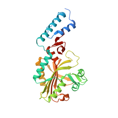
    Three for two: by using a Methanosarcina mazei PylRS triple mutant (Y306G, Y384F, I405R) the incorporation of two new exo-norbornene-containing pyrrolysine analogues was achieved. X-ray crystallographic analysis led to the identification of the crucial structural elements involved in substrate recognition by the evolved synthetase.


  • Organizational Affiliation

    Department of Chemistry, TU Munich, Lichtenbergstrasse 4, 85748 Garching (Germany). sabine.schneider@mytum.de.


Macromolecules
Find similar proteins by:  (by identity cutoff)  |  3D Structure
Entity ID: 1
MoleculeChains Sequence LengthOrganismDetailsImage
PYRROLYSINE--TRNA LIGASE291Methanosarcina mazeiMutation(s): 3 
EC: 6.1.1.26
UniProt
Find proteins for Q8PWY1 (Methanosarcina mazei (strain ATCC BAA-159 / DSM 3647 / Goe1 / Go1 / JCM 11833 / OCM 88))
Explore Q8PWY1 
Go to UniProtKB:  Q8PWY1
Entity Groups  
Sequence Clusters30% Identity50% Identity70% Identity90% Identity95% Identity100% Identity
UniProt GroupQ8PWY1
Sequence Annotations
Expand
  • Reference Sequence
Small Molecules
Ligands 4 Unique
IDChains Name / Formula / InChI Key2D Diagram3D Interactions
ANP
Query on ANP

Download Ideal Coordinates CCD File 
D [auth A]PHOSPHOAMINOPHOSPHONIC ACID-ADENYLATE ESTER
C10 H17 N6 O12 P3
PVKSNHVPLWYQGJ-KQYNXXCUSA-N
1PE
Query on 1PE

Download Ideal Coordinates CCD File 
E [auth A]PENTAETHYLENE GLYCOL
C10 H22 O6
JLFNLZLINWHATN-UHFFFAOYSA-N
EDO
Query on EDO

Download Ideal Coordinates CCD File 
F [auth A],
G [auth A],
H [auth A],
I [auth A]
1,2-ETHANEDIOL
C2 H6 O2
LYCAIKOWRPUZTN-UHFFFAOYSA-N
MG
Query on MG

Download Ideal Coordinates CCD File 
B [auth A],
C [auth A]
MAGNESIUM ION
Mg
JLVVSXFLKOJNIY-UHFFFAOYSA-N
Experimental Data & Validation

Experimental Data

  • Method: X-RAY DIFFRACTION
  • Resolution: 2.35 Å
  • R-Value Free: 0.204 
  • R-Value Work: 0.174 
  • R-Value Observed: 0.176 
  • Space Group: P 64
Unit Cell:
Length ( Å )Angle ( ˚ )
a = 105.742α = 90
b = 105.742β = 90
c = 69.674γ = 120
Software Package:
Software NamePurpose
REFMACrefinement
XDSdata reduction
SCALAdata scaling

Structure Validation

View Full Validation Report


Ligand Structure Quality Assessment 


Entry History 

Deposition Data

Revision History  (Full details and data files)

  • Version 1.0: 2013-07-31
    Type: Initial release
  • Version 1.1: 2013-09-25
    Changes: Database references
  • Version 1.2: 2013-12-25
    Changes: Database references
  • Version 1.3: 2023-12-20
    Changes: Data collection, Database references, Derived calculations, Other, Refinement description