4BUO

High Resolution Structure of Thermostable Agonist-bound Neurotensin Receptor 1 Mutant without Lysozyme Fusion


Experimental Data Snapshot

  • Method: X-RAY DIFFRACTION
  • Resolution: 2.75 Å
  • R-Value Free: 0.273 
  • R-Value Work: 0.248 
  • R-Value Observed: 0.249 

wwPDB Validation   3D Report Full Report


This is version 1.3 of the entry. See complete history


Literature

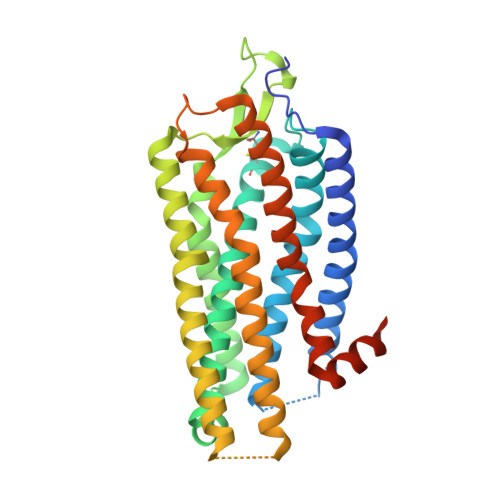
Structure of Signaling-Competent Neurotensin Receptor 1 Obtained by Directed Evolution in Escherichia Coli

Egloff, P.Hillenbrand, M.Klenk, C.Batyuk, A.Heine, P.Balada, S.Schlinkmann, K.M.Scott, D.J.Schuetz, M.Plueckthun, A.

(2014) Proc Natl Acad Sci U S A 111: E655

  • DOI: https://doi.org/10.1073/pnas.1317903111
  • Primary Citation of Related Structures:  
    3ZEV, 4BUO, 4BV0, 4BWB

  • PubMed Abstract: 

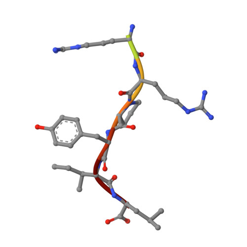
    Crystallography has advanced our understanding of G protein-coupled receptors, but low expression levels and instability in solution have limited structural insights to very few selected members of this large protein family. Using neurotensin receptor 1 (NTR1) as a proof of principle, we show that two directed evolution technologies that we recently developed have the potential to overcome these problems. We purified three neurotensin-bound NTR1 variants from Escherichia coli and determined their X-ray structures at up to 2.75 Å resolution using vapor diffusion crystallization experiments. A crystallized construct was pharmacologically characterized and exhibited ligand-dependent signaling, internalization, and wild-type-like agonist and antagonist affinities. Our structures are fully consistent with all biochemically defined ligand-contacting residues, and they represent an inactive NTR1 state at the cytosolic side. They exhibit significant differences to a previously determined NTR1 structure (Protein Data Bank ID code 4GRV) in the ligand-binding pocket and by the presence of the amphipathic helix 8. A comparison of helix 8 stability determinants between NTR1 and other crystallized G protein-coupled receptors suggests that the occupancy of the canonical position of the amphipathic helix is reduced to various extents in many receptors, and we have elucidated the sequence determinants for a stable helix 8. Our analysis also provides a structural rationale for the long-known effects of C-terminal palmitoylation reactions on G protein-coupled receptor signaling, receptor maturation, and desensitization.


  • Organizational Affiliation

    Department of Biochemistry, University of Zurich, 8057 Zurich, Switzerland.


Macromolecules
Find similar proteins by:  (by identity cutoff)  |  3D Structure
Entity ID: 1
MoleculeChains Sequence LengthOrganismDetailsImage
NEUROTENSIN RECEPTOR TYPE 1
A, B
335Rattus norvegicusMutation(s): 11 
Membrane Entity: Yes 
UniProt
Find proteins for P20789 (Rattus norvegicus)
Explore P20789 
Go to UniProtKB:  P20789
Entity Groups  
Sequence Clusters30% Identity50% Identity70% Identity90% Identity95% Identity100% Identity
UniProt GroupP20789
Sequence Annotations
Expand
  • Reference Sequence

Find similar proteins by:  Sequence   |   3D Structure  

Entity ID: 2
MoleculeChains Sequence LengthOrganismDetailsImage
NEUROTENSIN/NEUROMEDIN N
C, D
10Rattus norvegicusMutation(s): 0 
Membrane Entity: Yes 
UniProt
Find proteins for P20068 (Rattus norvegicus)
Explore P20068 
Go to UniProtKB:  P20068
Entity Groups  
Sequence Clusters30% Identity50% Identity70% Identity90% Identity95% Identity100% Identity
UniProt GroupP20068
Sequence Annotations
Expand
  • Reference Sequence
Small Molecules
Ligands 1 Unique
IDChains Name / Formula / InChI Key2D Diagram3D Interactions
GLY
Query on GLY

Download Ideal Coordinates CCD File 
E [auth A]
F [auth A]
G [auth A]
H [auth A]
I [auth A]
E [auth A],
F [auth A],
G [auth A],
H [auth A],
I [auth A],
J [auth A],
K [auth A],
L [auth A],
M [auth B],
N [auth B],
O [auth B]
GLYCINE
C2 H5 N O2
DHMQDGOQFOQNFH-UHFFFAOYSA-N
Experimental Data & Validation

Experimental Data

  • Method: X-RAY DIFFRACTION
  • Resolution: 2.75 Å
  • R-Value Free: 0.273 
  • R-Value Work: 0.248 
  • R-Value Observed: 0.249 
  • Space Group: P 21 21 21
Unit Cell:
Length ( Å )Angle ( ˚ )
a = 63.31α = 90
b = 89.4β = 90
c = 212.13γ = 90
Software Package:
Software NamePurpose
PHENIXrefinement
XDSdata reduction
XSCALEdata scaling
PHENIXphasing

Structure Validation

View Full Validation Report


Entry History 

Deposition Data

Revision History  (Full details and data files)

  • Version 1.0: 2014-01-29
    Type: Initial release
  • Version 1.1: 2014-02-05
    Changes: Database references
  • Version 1.2: 2014-02-26
    Changes: Database references
  • Version 1.3: 2023-12-20
    Changes: Data collection, Database references, Derived calculations, Other, Refinement description