4BTW

Crystal structure of human vascular adhesion protein-1 in complex with pyridazinone inhibitors


Experimental Data Snapshot

  • Method: X-RAY DIFFRACTION
  • Resolution: 2.80 Å
  • R-Value Free: 0.260 
  • R-Value Work: 0.205 
  • R-Value Observed: 0.208 

wwPDB Validation   3D Report Full Report


Ligand Structure Quality Assessment 


This is version 2.1 of the entry. See complete history


Literature

Novel Pyridazinone Inhibitors for Vascular Adhesion Protein- 1 (Vap-1): Old Target - New Inhibition Mode.

Bligt-Linden, E.Pihlavisto, M.Szatmari, I.Otwinowski, Z.Smith, D.J.Lazar, L.Fulop, F.Salminen, T.A.

(2013) J Med Chem 56: 9837

  • DOI: https://doi.org/10.1021/jm401372d
  • Primary Citation of Related Structures:  
    4BTW, 4BTX, 4BTY

  • PubMed Abstract: 

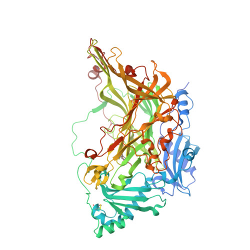
    Vascular adhesion protein-1 (VAP-1) is a primary amine oxidase and a drug target for inflammatory and vascular diseases. Despite extensive attempts to develop potent, specific, and reversible inhibitors of its enzyme activity, the task has proven challenging. Here we report the synthesis, inhibitory activity, and molecular binding mode of novel pyridazinone inhibitors, which show specificity for VAP-1 over monoamine and diamine oxidases. The crystal structures of three inhibitor-VAP-1 complexes show that these compounds bind reversibly into a unique binding site in the active site channel. Although they are good inhibitors of human VAP-1, they do not inhibit rodent VAP-1 well. To investigate this further, we used homology modeling and structural comparison to identify amino acid differences, which explain the species-specific binding properties. Our results prove the potency and specificity of these new inhibitors, and the detailed characterization of their binding mode is of importance for further development of VAP-1 inhibitors.


  • Organizational Affiliation

    Structural Bioinformatics Laboratory, Department of Biosciences, Åbo Akademi University , FI-20520 Turku, Finland.


Macromolecules
Find similar proteins by:  (by identity cutoff)  |  3D Structure
Entity ID: 1
MoleculeChains Sequence LengthOrganismDetailsImage
MEMBRANE PRIMARY AMINE OXIDASE
A, B
737Homo sapiensMutation(s): 0 
EC: 1.4.3.21
UniProt & NIH Common Fund Data Resources
Find proteins for Q16853 (Homo sapiens)
Explore Q16853 
Go to UniProtKB:  Q16853
PHAROS:  Q16853
GTEx:  ENSG00000131471 
Entity Groups  
Sequence Clusters30% Identity50% Identity70% Identity90% Identity95% Identity100% Identity
UniProt GroupQ16853
Sequence Annotations
Expand
  • Reference Sequence
Oligosaccharides

Help

Entity ID: 2
MoleculeChains Length2D Diagram Glycosylation3D Interactions
beta-D-mannopyranose-(1-4)-2-acetamido-2-deoxy-beta-D-glucopyranose-(1-4)-2-acetamido-2-deoxy-beta-D-glucopyranose
C
3N-Glycosylation
Glycosylation Resources
GlyTouCan:  G15407YE
GlyCosmos:  G15407YE
GlyGen:  G15407YE
Entity ID: 3
MoleculeChains Length2D Diagram Glycosylation3D Interactions
2-acetamido-2-deoxy-beta-D-glucopyranose-(1-4)-2-acetamido-2-deoxy-beta-D-glucopyranose
D, F
2N-Glycosylation
Glycosylation Resources
GlyTouCan:  G42666HT
GlyCosmos:  G42666HT
GlyGen:  G42666HT
Entity ID: 4
MoleculeChains Length2D Diagram Glycosylation3D Interactions
alpha-D-mannopyranose-(1-3)-[beta-D-mannopyranose-(1-6)]beta-D-mannopyranose-(1-4)-2-acetamido-2-deoxy-beta-D-glucopyranose-(1-4)-2-acetamido-2-deoxy-beta-D-glucopyranose
E
5N-Glycosylation
Glycosylation Resources
GlyTouCan:  G51945PF
GlyCosmos:  G51945PF
GlyGen:  G51945PF
Small Molecules
Ligands 4 Unique
IDChains Name / Formula / InChI Key2D Diagram3D Interactions
JW7
Query on JW7

Download Ideal Coordinates CCD File 
L [auth A],
T [auth B]
5-(cyclohexylamino)-2-phenyl-6-(1H-1,2,4-triazol-5-yl)-3(2H)-pyridazinone
C18 H20 N6 O
CRZJAUQQVQGZNE-UHFFFAOYSA-N
NAG
Query on NAG

Download Ideal Coordinates CCD File 
J [auth A]
K [auth A]
P [auth B]
Q [auth B]
R [auth B]
J [auth A],
K [auth A],
P [auth B],
Q [auth B],
R [auth B],
S [auth B]
2-acetamido-2-deoxy-beta-D-glucopyranose
C8 H15 N O6
OVRNDRQMDRJTHS-FMDGEEDCSA-N
CU
Query on CU

Download Ideal Coordinates CCD File 
G [auth A],
M [auth B]
COPPER (II) ION
Cu
JPVYNHNXODAKFH-UHFFFAOYSA-N
CA
Query on CA

Download Ideal Coordinates CCD File 
H [auth A],
I [auth A],
N [auth B],
O [auth B]
CALCIUM ION
Ca
BHPQYMZQTOCNFJ-UHFFFAOYSA-N
Modified Residues  1 Unique
IDChains TypeFormula2D DiagramParent
TPQ
Query on TPQ
A, B
L-PEPTIDE LINKINGC9 H9 N O5TYR
Binding Affinity Annotations 
IDSourceBinding Affinity
JW7 PDBBind:  4BTW IC50: 71 (nM) from 1 assay(s)
Experimental Data & Validation

Experimental Data

  • Method: X-RAY DIFFRACTION
  • Resolution: 2.80 Å
  • R-Value Free: 0.260 
  • R-Value Work: 0.205 
  • R-Value Observed: 0.208 
  • Space Group: P 65 2 2
Unit Cell:
Length ( Å )Angle ( ˚ )
a = 226.473α = 90
b = 226.473β = 90
c = 219.25γ = 120
Software Package:
Software NamePurpose
HKLdata reduction
HKLdata scaling
CCP4phasing
MOLREPphasing
REFMACrefinement

Structure Validation

View Full Validation Report


Ligand Structure Quality Assessment 


Entry History 

Deposition Data

Revision History  (Full details and data files)

  • Version 1.0: 2013-12-18
    Type: Initial release
  • Version 1.1: 2014-01-15
    Changes: Database references
  • Version 2.0: 2020-07-29
    Type: Remediation
    Reason: Carbohydrate remediation
    Changes: Advisory, Atomic model, Data collection, Derived calculations, Other, Structure summary
  • Version 2.1: 2023-12-20
    Changes: Data collection, Database references, Refinement description, Structure summary