


4BST
Structure of the ectodomain of LGR5 in complex with R-spondin-1 (Fu1Fu2) in P6122 crystal form
- PDB DOI: https://doi.org/10.2210/pdb4BST/pdb
- Classification: SIGNALING PROTEIN
- Organism(s): Homo sapiens
- Expression System: Homo sapiens
- Mutation(s): No
- Deposited: 2013-06-11 Released: 2013-06-19
Experimental Data Snapshot
- Method: X-RAY DIFFRACTION
- Resolution: 4.30 Å
- R-Value Free: 0.268
- R-Value Work: 0.243
- R-Value Observed: 0.244

This is version 2.0 of the entry. See complete history.

Macromolecules
Find similar proteins by:
(by identity cutoff) | 3D Structure
Entity ID: 1 | |||||
|---|---|---|---|---|---|
| Molecule | Chains | Sequence Length | Organism | Details | Image |
| LEUCINE-RICH REPEAT-CONTAINING G-PROTEIN COUPLED RECEPTOR 5 | 539 | Homo sapiens | Mutation(s): 0 | 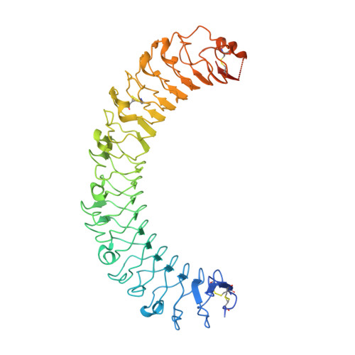 | |
UniProt & NIH Common Fund Data Resources | |||||
Find proteins for O75473 (Homo sapiens) Explore O75473 Go to UniProtKB: O75473 | |||||
PHAROS: O75473 GTEx: ENSG00000139292 | |||||
Entity Groups | |||||
| Sequence Clusters | 30% Identity50% Identity70% Identity90% Identity95% Identity100% Identity | ||||
| UniProt Group | O75473 | ||||
Sequence AnnotationsExpand | |||||
| |||||
Find similar proteins by:
(by identity cutoff) | 3D Structure
Entity ID: 2 | |||||
|---|---|---|---|---|---|
| Molecule | Chains | Sequence Length | Organism | Details | Image |
| R-SPONDIN-1 | 126 | Homo sapiens | Mutation(s): 0 | 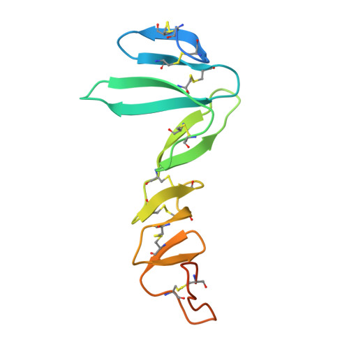 | |
UniProt & NIH Common Fund Data Resources | |||||
Find proteins for Q2MKA7 (Homo sapiens) Explore Q2MKA7 Go to UniProtKB: Q2MKA7 | |||||
PHAROS: Q2MKA7 GTEx: ENSG00000169218 | |||||
Entity Groups | |||||
| Sequence Clusters | 30% Identity50% Identity70% Identity90% Identity95% Identity100% Identity | ||||
| UniProt Group | Q2MKA7 | ||||
Sequence AnnotationsExpand | |||||
| |||||
Oligosaccharides

Small Molecules
| Ligands 1 Unique | |||||
|---|---|---|---|---|---|
| ID | Chains | Name / Formula / InChI Key | 2D Diagram | 3D Interactions | |
| NAG Query on NAG | F [auth A], G [auth A], H [auth A], I [auth B] | 2-acetamido-2-deoxy-beta-D-glucopyranose C8 H15 N O6 OVRNDRQMDRJTHS-FMDGEEDCSA-N |  | ||
Experimental Data & Validation
Experimental Data
- Method: X-RAY DIFFRACTION
- Resolution: 4.30 Å
- R-Value Free: 0.268
- R-Value Work: 0.243
- R-Value Observed: 0.244
- Space Group: P 61 2 2
Unit Cell:
| Length ( Å ) | Angle ( ˚ ) |
|---|---|
| a = 131.1 | α = 90 |
| b = 131.1 | β = 90 |
| c = 531.97 | γ = 120 |
| Software Name | Purpose |
|---|---|
| PHENIX | refinement |
Entry History
Deposition Data
- Released Date: 2013-06-19 Deposition Author(s): Peng, W.C., de Lau, W., Forneris, F., Granneman, J.C.M., Huch, M., Clevers, H., Gros, P.
Revision History (Full details and data files)
- Version 1.0: 2013-06-19
Type: Initial release - Version 1.1: 2013-07-24
Changes: Database references - Version 2.0: 2020-07-29
Type: Remediation
Reason: Carbohydrate remediation
Changes: Advisory, Atomic model, Data collection, Derived calculations, Other, Structure summary
