4BSK

Crystal structure of VEGF-C in complex with VEGFR-3 domains D1-2


Experimental Data Snapshot

  • Method: X-RAY DIFFRACTION
  • Resolution: 4.20 Å
  • R-Value Free: 0.372 
  • R-Value Work: 0.334 
  • R-Value Observed: 0.338 

wwPDB Validation   3D Report Full Report


Ligand Structure Quality Assessment 


This is version 3.1 of the entry. See complete history


Literature

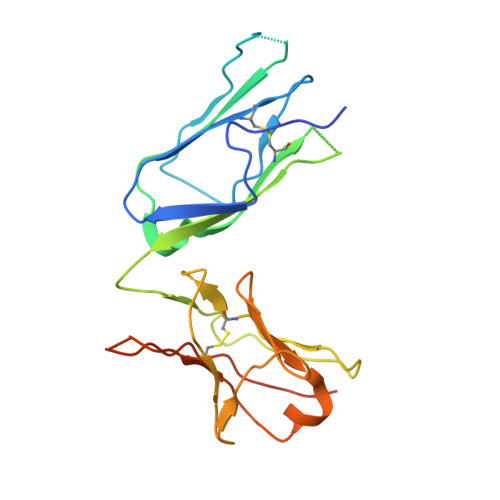
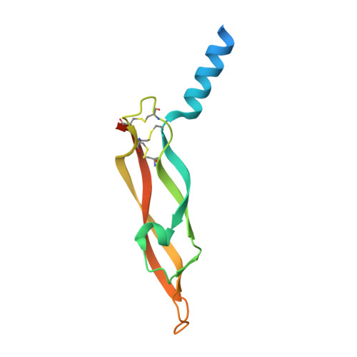
Structural and Mechanistic Insights Into Vegfr-3 Ligand Binding and Activation

Leppanen, V.-M.Tvorogov, D.Kisko, K.Prota, A.E.Jeltsch, M.Anisimov, A.Markovic-Mueller, S.Stuttfeld, E.Goldie, K.N.Ballmer-Hofer, K.Alitalo, K.

(2013) Proc Natl Acad Sci U S A 110: 12960

  • DOI: https://doi.org/10.1073/pnas.1301415110
  • Primary Citation of Related Structures:  
    4BSJ, 4BSK

  • PubMed Abstract: 

    Vascular endothelial growth factors (VEGFs) and their receptors (VEGFRs) are key drivers of blood and lymph vessel formation in development, but also in several pathological processes. VEGF-C signaling through VEGFR-3 promotes lymphangiogenesis, which is a clinically relevant target for treating lymphatic insufficiency and for blocking tumor angiogenesis and metastasis. The extracellular domain of VEGFRs consists of seven Ig homology domains; domains 1-3 (D1-3) are responsible for ligand binding, and the membrane-proximal domains 4-7 (D4-7) are involved in structural rearrangements essential for receptor dimerization and activation. Here we analyzed the crystal structures of VEGF-C in complex with VEGFR-3 domains D1-2 and of the VEGFR-3 D4-5 homodimer. The structures revealed a conserved ligand-binding interface in D2 and a unique mechanism for VEGFR dimerization and activation, with homotypic interactions in D5. Mutation of the conserved residues mediating the D5 interaction (Thr446 and Lys516) and the D7 interaction (Arg737) compromised VEGF-C induced VEGFR-3 activation. A thermodynamic analysis of VEGFR-3 deletion mutants showed that D3, D4-5, and D6-7 all contribute to ligand binding. A structural model of the VEGF-C/VEGFR-3 D1-7 complex derived from small-angle X-ray scattering data is consistent with the homotypic interactions in D5 and D7. Taken together, our data show that ligand-dependent homotypic interactions in D5 and D7 are essential for VEGFR activation, opening promising possibilities for the design of VEGFR-specific drugs.


  • Organizational Affiliation

    Translational Cancer Biology Program, Institute for Molecular Medicine Finland and Helsinki University Central Hospital, Biomedicum Helsinki, University of Helsinki, 00014 Helsinki, Finland.


Macromolecules
Find similar proteins by:  (by identity cutoff)  |  3D Structure
Entity ID: 1
MoleculeChains Sequence LengthOrganismDetailsImage
VASCULAR ENDOTHELIAL GROWTH FACTOR RECEPTOR 3214Homo sapiensMutation(s): 0 
EC: 2.7.10.1
UniProt & NIH Common Fund Data Resources
Find proteins for P35916 (Homo sapiens)
Explore P35916 
Go to UniProtKB:  P35916
PHAROS:  P35916
GTEx:  ENSG00000037280 
Entity Groups  
Sequence Clusters30% Identity50% Identity70% Identity90% Identity95% Identity100% Identity
UniProt GroupP35916
Sequence Annotations
Expand
  • Reference Sequence
Find similar proteins by:  (by identity cutoff)  |  3D Structure
Entity ID: 2
MoleculeChains Sequence LengthOrganismDetailsImage
VASCULAR ENDOTHELIAL GROWTH FACTOR CB [auth C]121Homo sapiensMutation(s): 1 
UniProt & NIH Common Fund Data Resources
Find proteins for P49767 (Homo sapiens)
Explore P49767 
Go to UniProtKB:  P49767
PHAROS:  P49767
GTEx:  ENSG00000150630 
Entity Groups  
Sequence Clusters30% Identity50% Identity70% Identity90% Identity95% Identity100% Identity
UniProt GroupP49767
Sequence Annotations
Expand
  • Reference Sequence
Oligosaccharides

Help

Entity ID: 3
MoleculeChains Length2D Diagram Glycosylation3D Interactions
2-acetamido-2-deoxy-beta-D-glucopyranose-(1-4)-2-acetamido-2-deoxy-beta-D-glucopyranoseC [auth B]2N-Glycosylation
Glycosylation Resources
GlyTouCan:  G42666HT
GlyCosmos:  G42666HT
GlyGen:  G42666HT
Experimental Data & Validation

Experimental Data

  • Method: X-RAY DIFFRACTION
  • Resolution: 4.20 Å
  • R-Value Free: 0.372 
  • R-Value Work: 0.334 
  • R-Value Observed: 0.338 
  • Space Group: I 2 3
Unit Cell:
Length ( Å )Angle ( ˚ )
a = 166.66α = 90
b = 166.66β = 90
c = 166.66γ = 90
Software Package:
Software NamePurpose
XDSdata reduction
XDSdata scaling
autoSHARPphasing
MOLREPphasing
PHENIXrefinement

Structure Validation

View Full Validation Report


Ligand Structure Quality Assessment 


Entry History 

Revision History  (Full details and data files)

  • Version 1.0: 2013-07-31
    Type: Initial release
  • Version 1.1: 2013-08-21
    Changes: Database references
  • Version 1.2: 2013-08-28
    Changes: Derived calculations
  • Version 2.0: 2018-04-11
    Changes: Advisory, Atomic model, Data collection, Derived calculations
  • Version 2.1: 2019-04-24
    Changes: Data collection, Source and taxonomy
  • Version 2.2: 2019-05-08
    Changes: Data collection, Experimental preparation
  • Version 3.0: 2020-07-29
    Type: Remediation
    Reason: Carbohydrate remediation
    Changes: Atomic model, Data collection, Derived calculations, Other, Structure summary
  • Version 3.1: 2023-12-20
    Changes: Data collection, Database references, Refinement description, Structure summary