4BQH

Crystal structure of the uridine diphosphate N-acetylglucosamine pyrophosphorylase from Trypanosoma brucei in complex with inhibitor


Experimental Data Snapshot

  • Method: X-RAY DIFFRACTION
  • Resolution: 1.75 Å
  • R-Value Free: 0.221 
  • R-Value Work: 0.189 
  • R-Value Observed: 0.190 

wwPDB Validation   3D Report Full Report


Ligand Structure Quality Assessment 


This is version 1.2 of the entry. See complete history


Literature

A Novel Allosteric Inhibitor of the Uridine Diphosphate N-Acetylglucosamine Pyrophosphorylase from Trypanosoma Brucei.

Urbaniak, M.D.Collie, I.T.Fang, W.Aristotelous, T.Eskilsson, S.Raimi, O.G.Harrison, J.Hopkins Navratolova, I.Frearson, J.A.Van Aalten, D.M.F.Ferguson, M.A.J.

(2013) ACS Chem Biol 8: 1981

  • DOI: https://doi.org/10.1021/cb400411x
  • Primary Citation of Related Structures:  
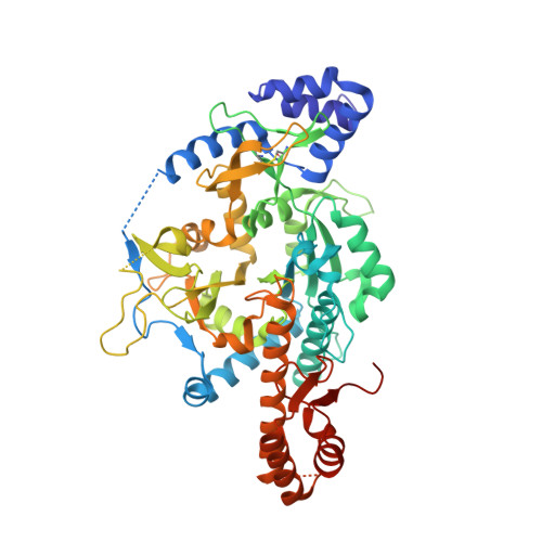
    4BQH

  • PubMed Abstract: 

    Uridine diphosphate N-acetylglucosamine pyrophosphorylase (UAP) catalyzes the final reaction in the biosynthesis of UDP-GlcNAc, an essential metabolite in many organisms including Trypanosoma brucei, the etiological agent of Human African Trypanosomiasis. High-throughput screening of recombinant T. brucei UAP identified a UTP-competitive inhibitor with selectivity over the human counterpart despite the high level of conservation of active site residues. Biophysical characterization of the UAP enzyme kinetics revealed that the human and trypanosome enzymes both display a strictly ordered bi-bi mechanism, but with the order of substrate binding reversed. Structural characterization of the T. brucei UAP-inhibitor complex revealed that the inhibitor binds at an allosteric site absent in the human homologue that prevents the conformational rearrangement required to bind UTP. The identification of a selective inhibitory allosteric binding site in the parasite enzyme has therapeutic potential.


  • Organizational Affiliation

    Division of Biological Chemistry and Drug Discovery, ‡Division of Molecular Microbiology, and §MRC Protein Phosphorylation and Ubiquitylation Unit, College of Life Sciences, University of Dundee , Dundee DD1 5EH, U.K.


Macromolecules
Find similar proteins by:  (by identity cutoff)  |  3D Structure
Entity ID: 1
MoleculeChains Sequence LengthOrganismDetailsImage
UDP-N-ACETYLGLUCOSAMINE PYROPHOSPHORYLASE549Trypanosoma bruceiMutation(s): 0 
EC: 2.7.7.23
UniProt
Find proteins for Q386Q8 (Trypanosoma brucei brucei (strain 927/4 GUTat10.1))
Explore Q386Q8 
Go to UniProtKB:  Q386Q8
Entity Groups  
Sequence Clusters30% Identity50% Identity70% Identity90% Identity95% Identity100% Identity
UniProt GroupQ386Q8
Sequence Annotations
Expand
  • Reference Sequence
Small Molecules
Ligands 2 Unique
IDChains Name / Formula / InChI Key2D Diagram3D Interactions
9VU
Query on 9VU

Download Ideal Coordinates CCD File 
C [auth A](3S)-3-[2-(1,3-benzodioxol-5-yl)-2-oxidanylidene-ethyl]-4-bromanyl-5-methyl-3-oxidanyl-1H-indol-2-one
C18 H14 Br N O5
SJXSNMXBXJIRLV-SFHVURJKSA-N
SO4
Query on SO4

Download Ideal Coordinates CCD File 
B [auth A],
D [auth A]
SULFATE ION
O4 S
QAOWNCQODCNURD-UHFFFAOYSA-L
Binding Affinity Annotations 
IDSourceBinding Affinity
9VU PDBBind:  4BQH Kd: 2580 (nM) from 1 assay(s)
Binding MOAD:  4BQH Kd: 2580 (nM) from 1 assay(s)
Experimental Data & Validation

Experimental Data

  • Method: X-RAY DIFFRACTION
  • Resolution: 1.75 Å
  • R-Value Free: 0.221 
  • R-Value Work: 0.189 
  • R-Value Observed: 0.190 
  • Space Group: C 2 2 21
Unit Cell:
Length ( Å )Angle ( ˚ )
a = 59.93α = 90
b = 103β = 90
c = 187.14γ = 90
Software Package:
Software NamePurpose
REFMACrefinement
iMOSFLMdata reduction
SCALEPACKdata scaling
BALBESphasing

Structure Validation

View Full Validation Report


Ligand Structure Quality Assessment 


Entry History 

Deposition Data

Revision History  (Full details and data files)

  • Version 1.0: 2013-07-17
    Type: Initial release
  • Version 1.1: 2013-10-09
    Changes: Database references
  • Version 1.2: 2023-12-20
    Changes: Data collection, Database references, Derived calculations, Other, Refinement description