4BKT

von Hippel Lindau protein:ElonginB:ElonginC complex, in complex with (2S,4R)-N-methyl-1-[2-(3-methyl-1,2-oxazol-5-yl)ethanoyl]-4-oxidanyl-pyrrolidine-2-carboxamide


Experimental Data Snapshot

  • Method: X-RAY DIFFRACTION
  • Resolution: 2.35 Å
  • R-Value Free: 0.228 
  • R-Value Work: 0.190 
  • R-Value Observed: 0.192 

wwPDB Validation   3D Report Full Report


Ligand Structure Quality Assessment 


This is version 1.2 of the entry. See complete history


Literature

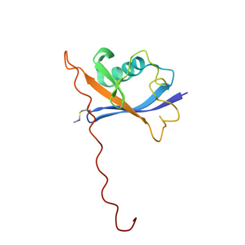
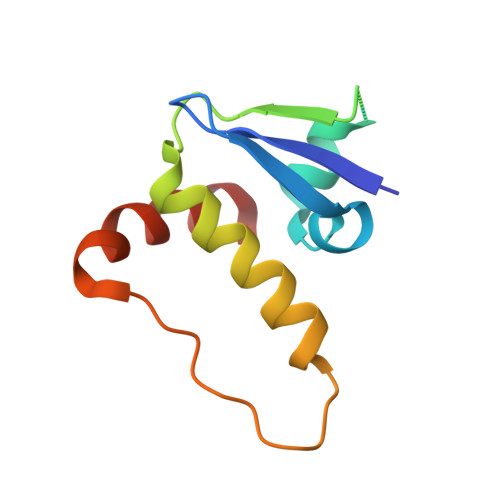
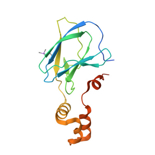
Is NMR Fragment Screening Fine-Tuned to Assess Druggability of Protein-Protein Interactions?

Dias, D.M.Van Molle, I.Baud, M.G.J.Galdeano, C.Geraldes, C.F.G.C.Ciulli, A.

(2014) ACS Med Chem Lett 5: 23

  • DOI: https://doi.org/10.1021/ml400296c
  • Primary Citation of Related Structures:  
    4BKS, 4BKT

  • PubMed Abstract: 

    Modulation of protein-protein interactions (PPIs) with small molecules has been hampered by a lack of lucid methods capable of reliably identifying high-quality hits. In fragment screening, the low ligand efficiencies associated with PPI target sites pose significant challenges to fragment binding detection. Here, we investigate the requirements for ligand-based NMR techniques to detect rule-of-three compliant fragments that form part of known high-affinity inhibitors of the PPI between the von Hippel-Lindau protein and the alpha subunit of hypoxia-inducible factor 1 (pVHL:HIF-1α). Careful triaging allowed rescuing weak but specific binding of fragments that would otherwise escape detection at this PPI. Further structural information provided by saturation transfer difference (STD) group epitope mapping, protein-based NMR, competitive isothermal titration calorimetry (ITC), and X-ray crystallography confirmed the binding mode of the rescued fragments. Our findings have important implications for PPI druggability assessment by fragment screening as they reveal an accessible threshold for fragment detection and validation.


  • Organizational Affiliation

    Department of Chemistry, University of Cambridge , Cambridge CB2 1EW, U.K. ; Department of Life Sciences, Faculty of Science and Technology, Centre for Neurosciences and Cell Biology and Chemistry Centre, University of Coimbra , Coimbra, Portugal.


Macromolecules
Find similar proteins by:  (by identity cutoff)  |  3D Structure
Entity ID: 1
MoleculeChains Sequence LengthOrganismDetailsImage
TRANSCRIPTION ELONGATION FACTOR B POLYPEPTIDE 2
A, D, G, J
104Homo sapiensMutation(s): 0 
UniProt & NIH Common Fund Data Resources
Find proteins for Q15370 (Homo sapiens)
Explore Q15370 
Go to UniProtKB:  Q15370
PHAROS:  Q15370
GTEx:  ENSG00000103363 
Entity Groups  
Sequence Clusters30% Identity50% Identity70% Identity90% Identity95% Identity100% Identity
UniProt GroupQ15370
Sequence Annotations
Expand
  • Reference Sequence
Find similar proteins by:  (by identity cutoff)  |  3D Structure
Entity ID: 2
MoleculeChains Sequence LengthOrganismDetailsImage
TRANSCRIPTION ELONGATION FACTOR B POLYPEPTIDE 1
B, E, H, K
97Homo sapiensMutation(s): 0 
UniProt & NIH Common Fund Data Resources
Find proteins for Q15369 (Homo sapiens)
Explore Q15369 
Go to UniProtKB:  Q15369
PHAROS:  Q15369
GTEx:  ENSG00000154582 
Entity Groups  
Sequence Clusters30% Identity50% Identity70% Identity90% Identity95% Identity100% Identity
UniProt GroupQ15369
Sequence Annotations
Expand
  • Reference Sequence
Find similar proteins by:  (by identity cutoff)  |  3D Structure
Entity ID: 3
MoleculeChains Sequence LengthOrganismDetailsImage
VON HIPPEL-LINDAU DISEASE TUMOR SUPPRESSOR
C, F, I, L
162Homo sapiensMutation(s): 0 
UniProt & NIH Common Fund Data Resources
Find proteins for P40337 (Homo sapiens)
Explore P40337 
Go to UniProtKB:  P40337
PHAROS:  P40337
GTEx:  ENSG00000134086 
Entity Groups  
Sequence Clusters30% Identity50% Identity70% Identity90% Identity95% Identity100% Identity
UniProt GroupP40337
Sequence Annotations
Expand
  • Reference Sequence
Small Molecules
Modified Residues  1 Unique
IDChains TypeFormula2D DiagramParent
CAS
Query on CAS
A, D, G, J
L-PEPTIDE LINKINGC5 H12 As N O2 SCYS
Binding Affinity Annotations 
IDSourceBinding Affinity
QD0 BindingDB:  4BKT Kd: 2.40e+5 (nM) from 1 assay(s)
PDBBind:  4BKT Kd: 2.40e+5 (nM) from 1 assay(s)
Binding MOAD:  4BKT Kd: 2.40e+5 (nM) from 1 assay(s)
Experimental Data & Validation

Experimental Data

  • Method: X-RAY DIFFRACTION
  • Resolution: 2.35 Å
  • R-Value Free: 0.228 
  • R-Value Work: 0.190 
  • R-Value Observed: 0.192 
  • Space Group: P 41 2 2
Unit Cell:
Length ( Å )Angle ( ˚ )
a = 93.18α = 90
b = 93.18β = 90
c = 364.5γ = 90
Software Package:
Software NamePurpose
BUSTERrefinement
XDSdata reduction
XSCALEdata scaling
BUSTERphasing

Structure Validation

View Full Validation Report


Ligand Structure Quality Assessment 


Entry History 

Deposition Data

Revision History  (Full details and data files)

  • Version 1.0: 2013-11-27
    Type: Initial release
  • Version 1.1: 2014-01-29
    Changes: Database references
  • Version 1.2: 2017-07-12
    Changes: Derived calculations