4BK7

Crystal Structure of a variant of the Major Birch Pollen Allergen Bet v 1


Experimental Data Snapshot

  • Method: X-RAY DIFFRACTION
  • Resolution: 1.14 Å
  • R-Value Free: 0.175 
  • R-Value Work: 0.134 
  • R-Value Observed: 0.137 

wwPDB Validation   3D Report Full Report


This is version 1.2 of the entry. See complete history


Literature

Stabilization of the Dimeric Birch Pollen Allergen Bet V 1 Impacts its Immunological Properties

Kofler, S.G.Ackaert, C.Samonig, M.Asam, C.Briza, P.Horeis-Hoeck, J.Cabrele, C.Ferreira, F.Duschl, A.Huber, C.Brandstetter, H.

(2014) J Biol Chem 289: 540

  • DOI: https://doi.org/10.1074/jbc.M113.518795
  • Primary Citation of Related Structures:  
    4BK6, 4BK7, 4BKC, 4BKD

  • PubMed Abstract: 

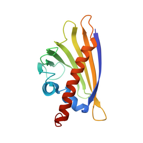
    Many allergens share several biophysical characteristics, including the capability to undergo oligomerization. The dimerization mechanism in Bet v 1 and its allergenic properties are so far poorly understood. Here, we report crystal structures of dimeric Bet v 1, revealing a noncanonical incorporation of cysteine at position 5 instead of genetically encoded tyrosine. Cysteine polysulfide bridging stabilized different dimeric assemblies, depending on the polysulfide linker length. These dimers represent quaternary arrangements that are frequently observed in related proteins, reflecting their prevalence in unmodified Bet v 1. These conclusions were corroborated by characteristic immunologic properties of monomeric and dimeric allergen variants. Hereby, residue 5 could be identified as an allergenic hot spot in Bet v 1. The presented results refine fundamental principles in protein chemistry and emphasize the importance of protein modifications in understanding the molecular basis of allergenicity.


  • Organizational Affiliation

    From the Structural Biology Group, Department of Molecular Biology.


Macromolecules
Find similar proteins by:  (by identity cutoff)  |  3D Structure
Entity ID: 1
MoleculeChains Sequence LengthOrganismDetailsImage
MAJOR POLLEN ALLERGEN BET V 1-A159Betula pendulaMutation(s): 1 
UniProt
Find proteins for P15494 (Betula pendula)
Explore P15494 
Go to UniProtKB:  P15494
Entity Groups  
Sequence Clusters30% Identity50% Identity70% Identity90% Identity95% Identity100% Identity
UniProt GroupP15494
Sequence Annotations
Expand
  • Reference Sequence
Small Molecules
Ligands 3 Unique
IDChains Name / Formula / InChI Key2D Diagram3D Interactions
MPD
Query on MPD

Download Ideal Coordinates CCD File 
G [auth A],
H [auth A],
I [auth A]
(4S)-2-METHYL-2,4-PENTANEDIOL
C6 H14 O2
SVTBMSDMJJWYQN-YFKPBYRVSA-N
SO4
Query on SO4

Download Ideal Coordinates CCD File 
B [auth A],
C [auth A],
D [auth A],
E [auth A],
F [auth A]
SULFATE ION
O4 S
QAOWNCQODCNURD-UHFFFAOYSA-L
NA
Query on NA

Download Ideal Coordinates CCD File 
J [auth A]SODIUM ION
Na
FKNQFGJONOIPTF-UHFFFAOYSA-N
Experimental Data & Validation

Experimental Data

  • Method: X-RAY DIFFRACTION
  • Resolution: 1.14 Å
  • R-Value Free: 0.175 
  • R-Value Work: 0.134 
  • R-Value Observed: 0.137 
  • Space Group: P 1 21 1
Unit Cell:
Length ( Å )Angle ( ˚ )
a = 32.732α = 90
b = 55.811β = 93.25
c = 38.099γ = 90
Software Package:
Software NamePurpose
REFMACrefinement
iMOSFLMdata reduction
SCALAdata scaling
PHASERphasing

Structure Validation

View Full Validation Report


Entry History 

Deposition Data

Revision History  (Full details and data files)

  • Version 1.0: 2013-11-27
    Type: Initial release
  • Version 1.1: 2014-01-15
    Changes: Database references
  • Version 1.2: 2023-12-20
    Changes: Data collection, Database references, Derived calculations, Other, Refinement description