4BJO

Nitrate in the active site of PTP1b is a putative mimetic of the transition state


Experimental Data Snapshot

  • Method: X-RAY DIFFRACTION
  • Resolution: 2.06 Å
  • R-Value Free: 0.239 
  • R-Value Work: 0.215 
  • R-Value Observed: 0.217 

wwPDB Validation   3D Report Full Report


This is version 1.3 of the entry. See complete history


Literature

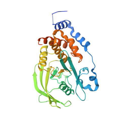
Nitrate in the Active Site of Protein Tyrosine Phosphatase 1B is a Putative Mimetic of the Transition State.

Kenny, P.W.Newman, J.Peat, T.S.

(2014) Acta Crystallogr D Biol Crystallogr 70: 565

  • DOI: https://doi.org/10.1107/S1399004713031052
  • Primary Citation of Related Structures:  
    4BJO

  • PubMed Abstract: 

    The X-ray crystal structure of the complex of protein tyrosine phosphatase 1B with nitrate anion has been determined and modelled quantum-mechanically. Two protomers were present in the structure, one with the mechanistically important WPD loop closed and the other with this loop open. Nitrate was observed bound to each protomer, making close contacts with the S atom of the catalytic cysteine and a tyrosine residue from a crystallographically related protomer.


  • Organizational Affiliation

    CSIRO Materials, Science and Engineering, 343 Royal Parade, Parkville, VIC 3052, Australia.


Macromolecules
Find similar proteins by:  (by identity cutoff)  |  3D Structure
Entity ID: 1
MoleculeChains Sequence LengthOrganismDetailsImage
TYROSINE-PROTEIN PHOSPHATASE NON-RECEPTOR TYPE 1
A, B
338Homo sapiensMutation(s): 0 
EC: 3.1.3.48
UniProt & NIH Common Fund Data Resources
Find proteins for P18031 (Homo sapiens)
Explore P18031 
Go to UniProtKB:  P18031
PHAROS:  P18031
GTEx:  ENSG00000196396 
Entity Groups  
Sequence Clusters30% Identity50% Identity70% Identity90% Identity95% Identity100% Identity
UniProt GroupP18031
Sequence Annotations
Expand
  • Reference Sequence
Small Molecules
Ligands 4 Unique
IDChains Name / Formula / InChI Key2D Diagram3D Interactions
EDO
Query on EDO

Download Ideal Coordinates CCD File 
G [auth A]1,2-ETHANEDIOL
C2 H6 O2
LYCAIKOWRPUZTN-UHFFFAOYSA-N
NO3
Query on NO3

Download Ideal Coordinates CCD File 
F [auth A],
K [auth B]
NITRATE ION
N O3
NHNBFGGVMKEFGY-UHFFFAOYSA-N
CL
Query on CL

Download Ideal Coordinates CCD File 
C [auth A],
D [auth A],
I [auth B],
J [auth B]
CHLORIDE ION
Cl
VEXZGXHMUGYJMC-UHFFFAOYSA-M
MG
Query on MG

Download Ideal Coordinates CCD File 
E [auth A],
H [auth A]
MAGNESIUM ION
Mg
JLVVSXFLKOJNIY-UHFFFAOYSA-N
Experimental Data & Validation

Experimental Data

  • Method: X-RAY DIFFRACTION
  • Resolution: 2.06 Å
  • R-Value Free: 0.239 
  • R-Value Work: 0.215 
  • R-Value Observed: 0.217 
  • Space Group: C 1 2 1
Unit Cell:
Length ( Å )Angle ( ˚ )
a = 161.62α = 90
b = 66.765β = 95.01
c = 68.771γ = 90
Software Package:
Software NamePurpose
REFMACrefinement
XDSdata reduction
SCALAdata scaling
PHASERphasing

Structure Validation

View Full Validation Report


Entry History 

Deposition Data

Revision History  (Full details and data files)

  • Version 1.0: 2014-02-12
    Type: Initial release
  • Version 1.1: 2014-02-26
    Changes: Database references
  • Version 1.2: 2019-05-08
    Changes: Data collection, Experimental preparation, Other
  • Version 1.3: 2023-12-20
    Changes: Data collection, Database references, Derived calculations, Other, Refinement description